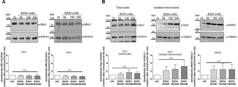
Figure 3. Protein expression of mitochondria-shaping proteins is not severely affected by 24 hours of ethanol exposure in human PCLS.
(A) Fusion pathway in PCLS treated with ethanol (EtOH). The protein expression of Mitofusin-1 (Mfn1) and Mitofusin-2 (Mfn2) was the same in untreated slices and after 24 hours of ethanol at the indicated doses (P>0.05).
(B) Fragmentation pathway in PCLS treated with ethanol (EtOH). The protein expression of Dynamin-1-like protein (Drp1) and Mitochondrial dynamics protein of 51KDa (MiD51), and the localisation of Drp1 on mitochondria was analysed by Western Blot in PCLS treated with the indicated doses of EtOH (P>0.05).
In all panels representative western blots are shown along with the densitometric analysis (mean ± SEM, n>3).

