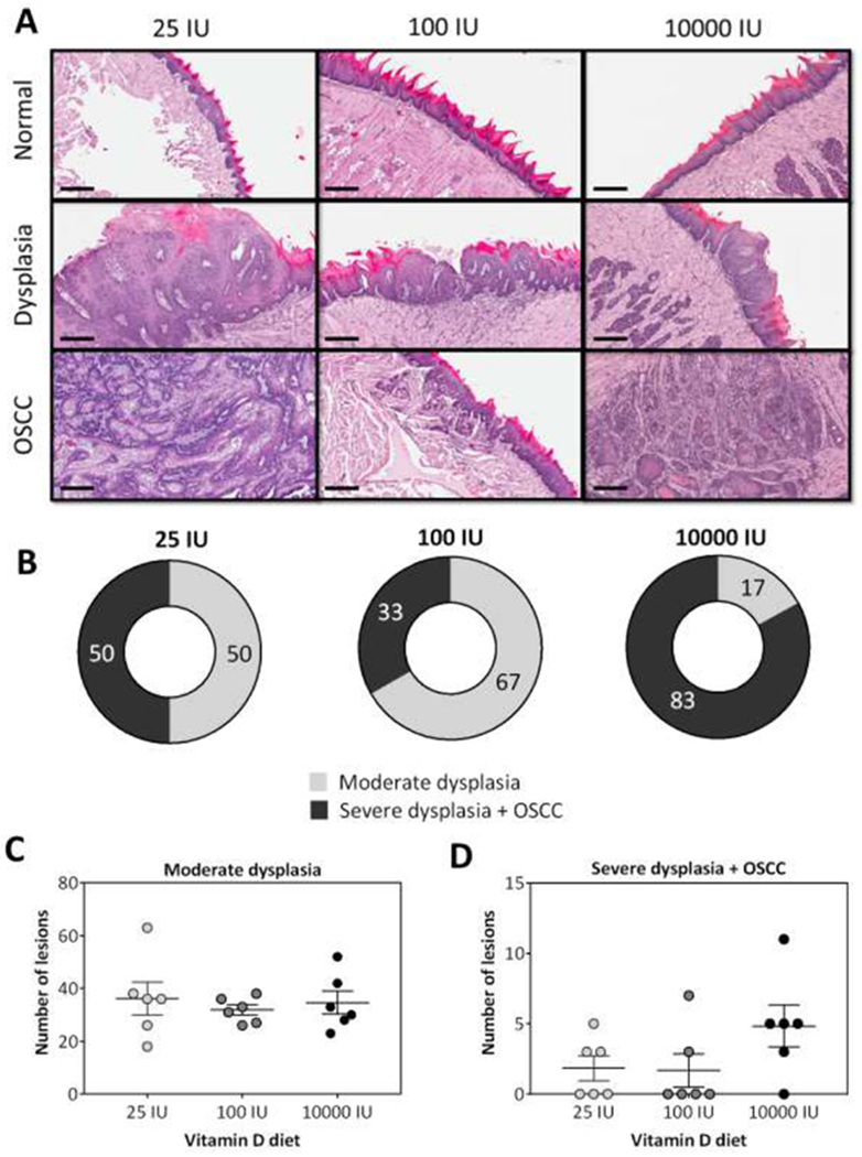
Figure 2. Impact of dietary vitamin D3 intake on incidence and multiplicity of oral premalignant lesions and OSCC.

(A) Photomicrographs of hematoxylin and eosin (H&E) stained sections (10X, scale bar - 200μm) of normal tongue epithelium, dysplasia and OSCC of a representative mouse from each diet group. (B) Donut charts illustrate the incidence (%) of oral premalignant and malignant lesions in mice maintained on the three diets. The number of oral lesions (Moderate dysplasia; C) (Severe dysplasia + OSCC; D) across the three diet groups are also shown (p>0.05). Histologic evaluation revealed lowest incidence of severe dysplasia + OSCC in mice maintained on the adequate diet while mice maintained on 10000 IU showed the highest incidence of OSCC. Reported values represent mean ± standard error.
