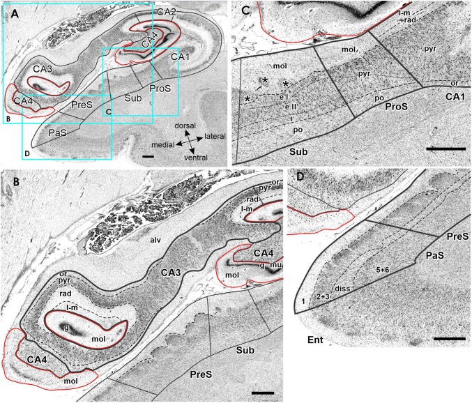
Fig. 4.
Cytoarchitecture of the hippocampal head and adjacent subicular complex. a Overview of an exemplary section comparable in its rostro-caudal position to section 3961 in Fig. 3. Cutouts (position highlighted by blue frames in a demonstrate differences in cytoarchitecture between b hippocampal regions FD, CA4 and CA3, c CA1, ProS and Sub, as well as d PreS and PaS. The dotted line in ProS highlights the gradual decrease in the width of the outer pyramidal sublayer (which contains pyramids typical of the CA region) and concomitant increase in the width of the inner pyramidal sublayer (which contains typical subicular pyramids). The dotted lines in Sub separate the two external sublayers (e I and e II) of the pyramidal layer from its internal (i) sublayer. Asterisks highlight the clusters of layer 2 cells from PreS which invade Sub. 1 layer 1 (molecular layer), 2 + 3 layers 2 and 3 (pyramidal layers), 5 + 6 layers 5 (parvocellular layer) and 6 (polymorph layer), alv alveum, CA1–CA4 regions 1–4 of the cornu Ammonis, diss dissecans layer, e I external sublayer I of the pyramidal layer, e II external sublayer II of the pyramidal layer, Ent entorhinal cortex, FD fascia dentata, g granular layer, i internal sublayer of the pyramidal layer, l-m lacunosum-molecular layer, mol molecular layer, mu multiform layer, or oriens layer, PaS parasubiculum, po polymorph layer, PreS presubiculum, ProS prosubiculum, pyr pyramidal layer, rad radiatum layer. Scale bar 1 mm

