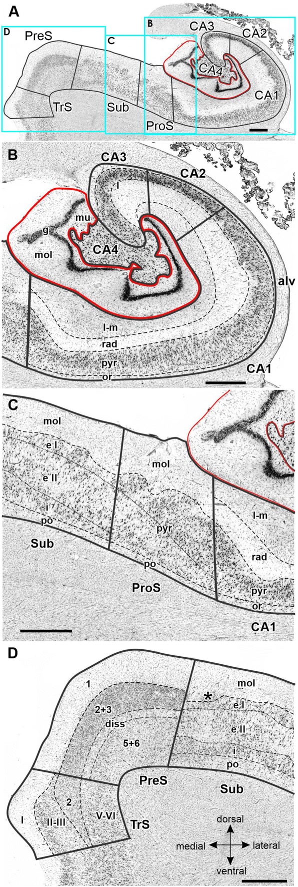
Fig. 5.

Cytoarchitecture of the hippocampal body and adjacent subicular complex. a Overview of an exemplary section comparable to the sectioning level 3241 in Fig. 3. Cutouts (position highlighted by blue frames in a demonstrate differences in cytoarchitecture between b hippocampal regions FD, CA4, CA3, CA2 and CA1, d subicular regions Sub, PreS and TrS. Asterisk highlights a cluster of layer 2 cells from PreS which invades Sub. Roman numerals indicate isocortical layers. 1 layer 1 (molecular layer), 2 + 3 layers 2 and 3 (pyramidal layers), 5 + 6 layers 5 (parvocellular layer) and 6 (polymorph layer), alv alveum, CA1–CA4 regions 1–4 of the cornu Ammonis, diss dissecans layer, g granular layer, l lucidum layer (note, that its border with the radiatum layer has not been indicated, because it is not revealed by the silver cell body staining), l-m lacunosum-molecular layer, mol molecular layer, mu multiform layer, or oriens layer, po polymorph layer, PreS presubiculum, ProS prosubiculum, pyr pyramidal layer, rad radiatum layer, Sub subiculum, TrS transsubiculum. Scale bars 1 mm
