
Fig. 9.
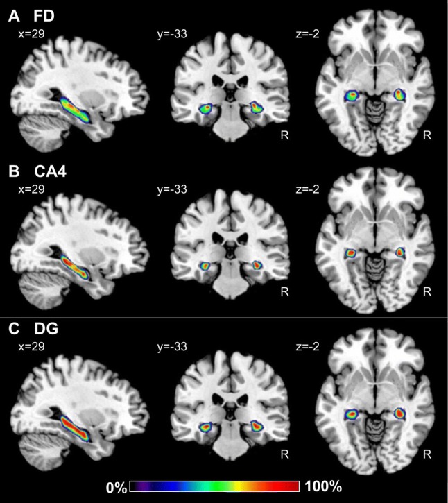
Continuous probabilistic maps of the cytoarchitectonically identifiable fascia dentata (FD, a) and CA4 region (b), as well as of the macroscopically identifiable dentate gyrus (DG, c) hippocampal and subicular regions overlayed on sagittal, coronal, and horizontal sections of the MNI single subject template (Evans et al. 2012). Stereotaxic coordinates are given in anatomical MNI space (Amunts et al. 2005). Note, that the novel workflows used for the computation of probabilistic maps result in a better reconstruction of the DG than in the previously published version of this map (compare c with figure 3 of Amunts et al. 2005). Color bar reflects probability of a region in a particular voxel. R right hemisphere