
Figure 4.
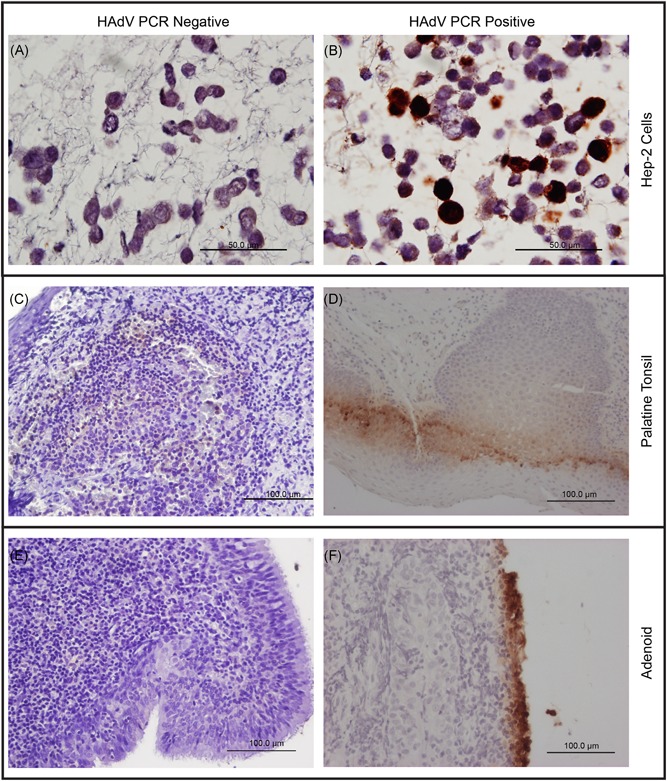
Immunohistochemistry for HAdV of noninfected and infected Hep‐2 cells, palatine tonsils and adenoids from patients with adenotonsillar chronic diseases. A, Noninfected Hep‐2 cells as negative controls counterstained with Hematoxylin and Eosin. B, HAdV‐infected Hep‐2 cells as positive controls counterstained with Hematoxylin and Eosin. C, Representative palatine tonsil from a patient without HAdV detectable by qPCR. D, Representative palatine tonsil from an HAdV‐positive patient. E, Representative adenoid from a patient without HAdV detectable by qPCR. F, Representative adenoid from a patient with HAdV detectable by qPCR, illustrating the presence of viral antigens in superficial cells. The positive signal is visible as brown color. The adenoids and palatine tonsils shown here were obtained from the same patient. HAdV, human adenovirus; qPCR, real‐time PCR