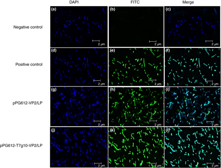
Figure 1.

Confocal laser scanning microscopic detection of surface expression of VP2 protein. Our results illustrate that there is a green fluorescent signal on the cell surface of strains pPG612‐VP2/LP, pPG612‐T7g10‐VP2/LP and positive control but no signals on negative control (pPG612/LP), indicating that the protein of interest is expressed and displayed successfully on the surface of recombinant Lactobacillus. a, b, c: pPG612/LP (negative control); d, e, f: pPG612‐VP2/Lactobacillus casei 393 (positive control); g, h, i: pPG612‐VP2/LP; j, k, l: pPG612‐T7g10‐VP2/LP. [Colour figure can be viewed at http://wileyonlinelibrary.com]
