
Figure 6.
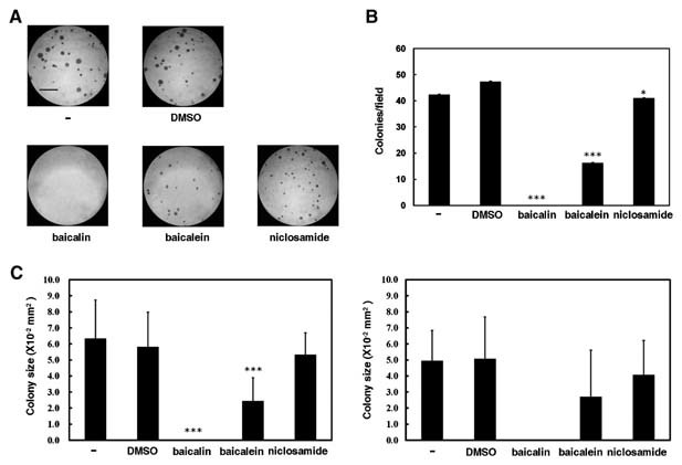
Colony‐forming ability of K562 cells is suppressed by baicalin and baicalein, but not by niclosamide. A: In the presence of an equal volume of PBS (−), DMSO, 100 µM baicalin, 10 µM baicalein, and 1.25 µM niclosamide, K562 cells were mixed with top agar in 6‐well plates. Cells were incubated for 7 days and then the plates were stained with 0.5 ml of 0.005% crystal violet for 1 h. Scale bar: 1.0 mm. Colonies larger than 0.1 mm in diameter were counted and mean colony sizes were estimated under microscope from 10 random fields. B: Data of colony numbers are representative of mean values and standard deviations from three independent experiments. C: Data of mean colony sizes are shown from one experiment (left) or mean values and standard deviations from three independent experiments (right). Because variation of colony sizes was very high, the change of mean colony sizes from three independent experiments is not statistically significant. Therefore, mean colony sizes from one experiment were also shown. *P < 0.05; ***P < 0.001 compared with vehicle.