
Figure 4.
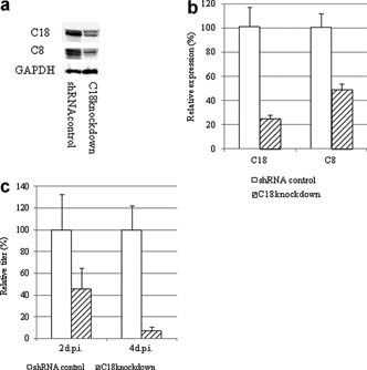
a and b: Cloning of C18 knocked‐down and shRNA control A549 cells. a: Western blot analysis of C8/C18 expression. Cells were mixed with SDS sample buffer and separated by 15% SDS–PAGE. C18 was detected with a biotin‐conjugated mouse monoclonal antibody (DC‐10; Abcam) and HRP‐conjugated streptavidin (Zymed). C8 was detected using a mouse monoclonal antibody (GeneTex) and HRP‐conjugated goat anti‐mouse antibodies (Rockland, Gilbertsville, PA). GAPDH was detected with rabbit polyclonal antibodies (Imgenex, San Diego, CA) and HRP‐conjugated guinea pig anti‐rabbit antibodies (Rockland). b: C8 and C18 mRNA expression in cloned cells. Total RNA was extracted, and first‐strand cDNA was synthesized for real‐time PCR as described in the legend for Figure 1 using GAPDH as an internal control (n = 7; *P < 0.01). c: Viral replication kinetics in C18 knocked‐down A549 cells. Cells were infected with RSV at an MOI of 1 and incubated for 2–4 days. Cells were collected and the titers were determined by plaque assay using HEp‐2 cells (n = 4).