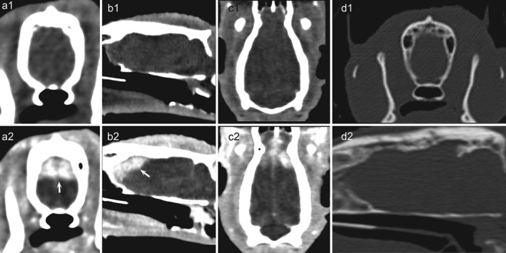
Figure 1.

CT‐examination of the head (120 kVp, 100 mA, slice thickness 0 · 5 mm, FOV 180). Transverse (a1, a2), sagittal (b1, b2) and dorsal (c1, c2) reconstructions displayed with a soft tissue window show an isodense pre‐contrast area with a strong post‐contrast enhancement (arrows) in the dorsal part of the right and left forebrain. The lesion shows a wide basis with a large osseous contact. No underlying bone lysis or sclerosis is observed (d1, d2: transverse and sagittal reconstructions displayed with a bone window)
