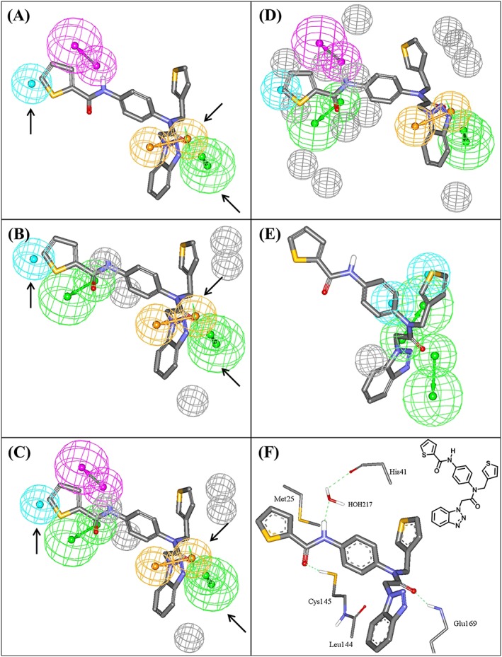
Figure 3.

Pharmacophoric features of the QSAR‐guided pharmacophores and the corresponding merged model: green‐vectored spheres: HBA; blue spheres: Hbic; purple‐vectored spheres: HBD; and orange‐vectored spheres: RingArom, (A) Hypo(N‐T1‐1), (B) Hypo(K‐T5‐3), (C) Merged‐Hypo(K‐T5‐3/N‐T1‐1), (D) Refined Merged‐Hypo(K‐T5‐3/N‐T1‐1), and (E) Hypo(L‐T5‐2) fitted against co‐crystallized ligand within HKU4‐CoV 3CLpro (compound 1, IC50 = 0.33μM, PDB code 4YOI, 1.8 Ǻ). (F) Ligand co‐crystallized within HKU4‐CoV 3CLpro and the chemical structure of the co‐crystallized ligand. Arrows point to closely positioned common features in Hypo(N‐T1‐1) and Hypo(K‐T5‐3) allowing for merging. The 3D coordinates of these pharmacophores are shown in Table S6. HBA, hydrogen bond acceptor; HBD, hydrogen bond donor
