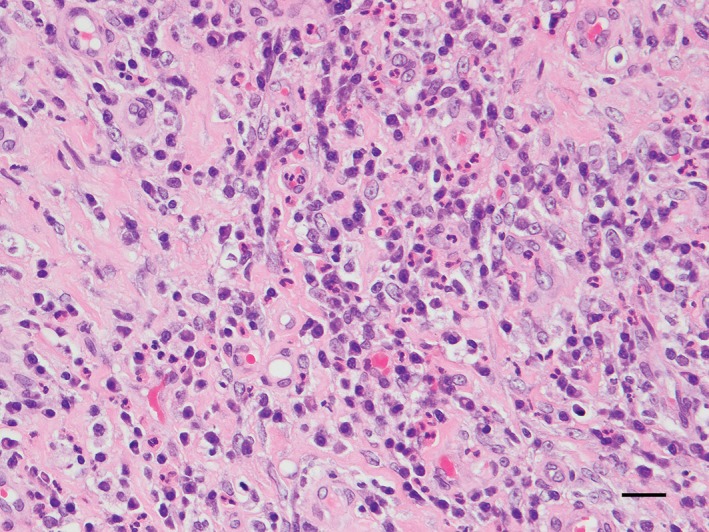
Figure 3.

Photomicrograph of the mesenteric nodules. The foci were composed of thin trabeculae of dense collagen fibres mixed with fibroblasts, numerous eosinophils and fewer lymphocytes, plasma cells and histiocytes. Haematoxylin and eosin stain, bar=20 μm
