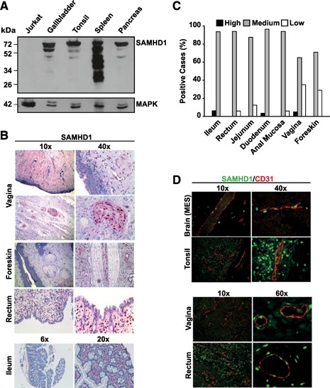
Figure 2.

SAMHD1 is abundantly expressed in solid organs, gut‐associated lymphoid tissue, at mucosal sites of anogenital HIV transmission, and in CD31+ vascular endothelial cells in vivo. (A) Western blot analysis of SAMHD1 in homogenates from selected human tissues (gallbladder, tonsil, spleen, and pancreas) and SAMHD1‐negative Jurkat‐TAg cells. Cell lysates were separated by SDS‐PAGE, and nitrocellulose membranes were probed with a polyclonal rabbit anti‐SAMHD1 antiserum. MAPK: loading control. (B) Stainings of ileum, rectal, foreskin, and vaginal tissues were performed with a polyclonal anti‐SAMHD1 antibody, followed by incubation with secondary antibody solutions and substrate development with Permanent Red. Nuclei were counterstained with hematoxylin. (C) Rating of SAMHD1 expression in GALT and HIV transmission‐site tissues, according to the scoring system used in Fig. 1C. Histogram bars depict the percentage of cases with high (black), medium (gray), or low (open) expression ratings. The percentage of samples with negative ratings is not shown. (D) Immunofluorescence coexpression analysis of SAMHD1 (green nuclear staining) and vessel endothelium (CD31+, red membranous, and cytoplasmic staining) in selected human tissues. Shown are 2‐channel merged images. MES, Mesencephalon.
