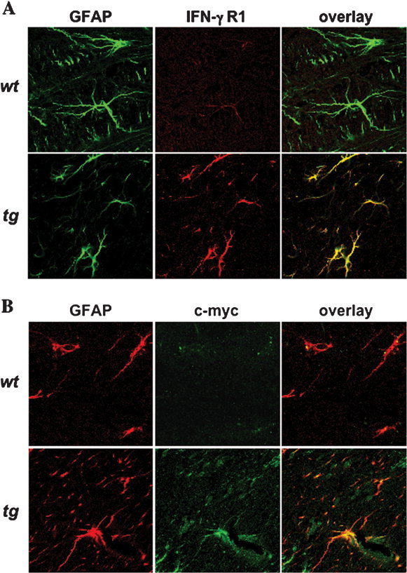
Figure 4.

Expression of the tg IFN‐γR1 by astrocytes. IFN‐γR1 (A), c‐myc (B), and astrocyte‐specific expression analyzed by confocal microscopy. Control wt (top) and tg (bottom) mice were examined for IFN‐γR1 expression (A, red) and GFAP (green). Expression of the transgene was confirmed by expression of c‐myc (B, green) and GFAP (red). No evidence for ectopic expression was detected in other CNS cell types in tg or wt mice.
