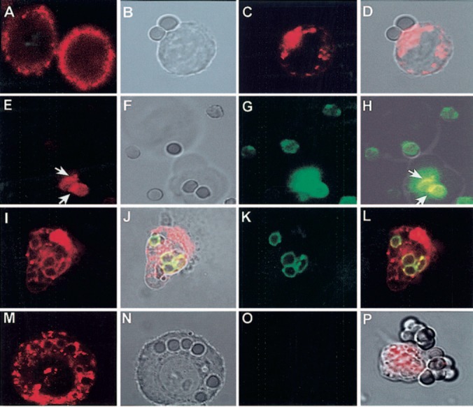
Figure 1.

CD13 redistributes to the phagocytic cup during FcγR‐mediated phagocytosis in U‐937 cells. (A) CD13 distribution on resting cells. (B–D) U‐937 cells were incubated with IgG‐opsonized erythrocytes for 60 min at 37°C. Cells were fixed, and CD13 was detected by incubation with TR‐labeled F(ab)′2 fragments of mAb anti‐CD13. (D) Redistributed CD13 in the zone of contact with erythrocytes. (E–H) U‐937 cells were incubated with FITC‐IgG‐opsonized erythrocytes for 60 min at 37°C. Cells were chilled, and CD13 was detected by incubation with TR‐labeled F(ab)′2 fragments of mAb anti‐CD13. CD13 is visualized in the zones of the membrane in contact with erythrocytes (E, white arrows). Cell membranes are out of focus (see transmitted light, F), impeding detection of distribution of the rest of the population of CD13 molecules in this slice. Erythrocytes are fluorescent in the green channel (G). (H) Projection shows in yellow sites of colocalization of FcγR (IgG‐opsonized erythrocytes) and CD13 (arrows). (I–L) U‐937 cells were incubated with TR‐labeled F(ab)′2 fragments of anti‐CD13 mAb at 4°C before phagocytosis, washed, and incubated with FITC‐IgG‐opsonized erythrocytes for 90 min at 37°C. Noninternalized erythrocytes were lysed before observation in the confocal microscope. Erythrocytes internalized into phagosomes (J, K) are surrounded by CD13 (I). (J and L) Colocalization of the CD13‐red signal with FITC‐IgG‐labeled erythrocytes in the phagosomes. (M–O) Similar experiment as that shown in I–L but with erythrocytes opsonized with nonlabeled IgG. CD13 is seen inside the phagosomes (M). No fluorescence is seen in the green channel (O). (P) Transmitted light and red channel‐composed image of U‐937 cells incubated with TR‐labeled F(ab)′2 fragments of anti‐CD13 mAb before incubation with IgG‐opsonized erythrocytes for 60 min at 4°C. CD13 does not redistribute to zones of contact with erythrocytes.
