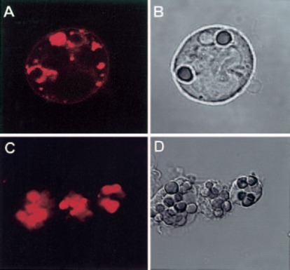
Figure 2.

CD13 redistributes to the phagocytic cup during FcγR‐mediated phagocytosis in peripheral blood human monocytes. (A, B) Human monocytes were incubated with TR‐labeled F(ab)′2 fragments of anti‐CD13 mAb at 4°C before phagocytosis, washed, and incubated with IgG‐opsonized erythrocytes for 15 min at 37°C. Noninternalized erythrocytes were lysed before observation in the confocal microscope. Erythrocytes internalized in phagosomes are surrounded by CD13. Distribution of CD13 label in the phagosomes is visualized as a uniform red staining in one phagosome and as a ring surrounding the other two internalized erythrocytes as a result of the plane of the X‐Y slice. (C, D) After 30 min, phagosomes show internalized CD13.
