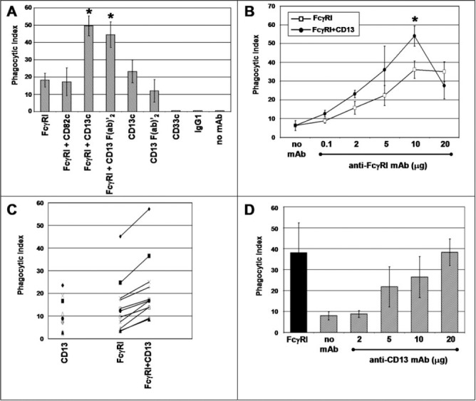
Figure 3.

Coaggregation of FcγRI with CD13 increases the phagocytic capacity of U‐937 cells and peripheral blood human monocytes. (A) U‐937 cells were incubated with 10 μg of the mAb specific for the indicated molecules for 30 min at 4°C. After washing, the cells were incubated for 90 min at 37°C with erythrocytes opsonized with F(ab)′2 fragments of biotin‐labeled goat anti‐mouse IgG (EBS‐Ab). After lysis of noninternalized erythrocytes, phagocytosis was evaluated by light microscopy. Data are presented as the number of EBS‐Ab internalized per 100 cells (Phagocytic Index). Mean ± sd of six independent experiments; c = complete mAb. (B) Peripheral blood monocytes from human donors were incubated with the indicated concentrations of anti‐FcγRI mAb with (•) or without (□) F(ab)′2 fragments of anti‐CD13 mAb (10 μg) for 30 min. After washing, incubation with EBS‐Ab was carried out at 37°C for 30 min. After lysis of noninternalized erythrocytes, the phagocytic index was determined by light microscopy. Data are the mean phagocytic index (±se) of at least four different donors for each point. (∗) Statistically significant difference comparing FcγRI + CD13 with FcγRI alone; P < 0.05. (C) Peripheral blood monocytes from different human donors were incubated with 10 μg anti‐CD13 mAb, 10 μg anti‐FcγRI, or the combination of the two antibodies and subsequently, with EBS‐Ab, as indicated above. Each distinct symbol represents a different donor. (D) Peripheral blood monocytes from different human donors were incubated with different concentrations of anti‐CD13 452 mAb and subsequently with EBS‐Ab. Data represent mean and sdof at least four donors. Solid bar represents the phagocytic index obtained with 10 μg anti‐FcγRI mAb.
