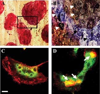
Figure 1.

Gigantic osteoclasts formed in vitro under acidic culture conditions. A: Low power transmitted light micrograph of TRAP‐stained osteoclasts on a bone slice formed during culture from day 7 to 14 under acid conditions (pH 6.8) showing extreme size (approximately 2 mm diameter) (numerous nuclei, >200, are present but obscured by the intense TRAP staining) (mag. 5×). B: Higher power reflected light micrograph of combined TRAP‐ and toluidine blue‐stained osteoclasts; detail of the area marked in (A) showing numerous resorption lacunae under and associated with two osteoclasts (examples are identified by arrows) (mag. 10×). C,D: Immunostaining of osteoclasts for cathepsin K formed under control (pH 7.4) (C) and acidic (pH 6.8) conditions (D). F‐actin rings are indicated by arrows in (D). Scale bar (in C) = 20 µm. These cells are representative examples used for the analyses summarized in Table 1.
