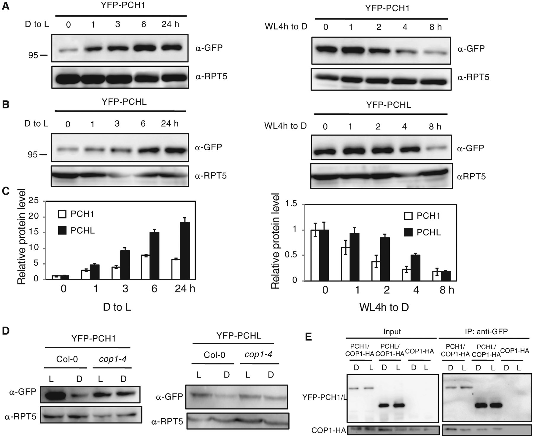
Figure 5. PCH1 and PCHL Are Unstable in the Dark and Their Degradation Is Mediated by COP1.

(A and B) Western blot analyses of PCH1 (A) and PCHL (B) protein (by GFP antibody) in PCH1OE and PCHLOE plants. The plants grown on MS were first illuminated for 4 h to induce germination. Four-day-old dark-grown seedlings were exposed to light for up to 24 h (both left panels) or first incubated in the light for 4 h and then left in darkness up to 8 h (both right panels). RPT5 was detected as loading control. Experiments were repeated three times with the same results, and a representative experiment is shown.
(C) Bar graph shows quantitative data from the western blots shown above. Error bars indicate SEM (n = 3 biological repeats).
(D) Western blots show that PCH1 (left) and PCHL (right) are degraded slower in cop1–4 mutants compared with wild-type background. Four-day-old dark-grown seedlings were illuminated for 4 h (L) or first illuminated for 4 h and then incubated in the dark for another 4 h (D).
(E)YFP-PCH1/PCHL interacts with COP1-HA in vivo. The input and pellet fractions are indicated. Co-IP was carried out using the anti-GFP antibody and then probed with anti-GFP and anti-HA antibodies.
