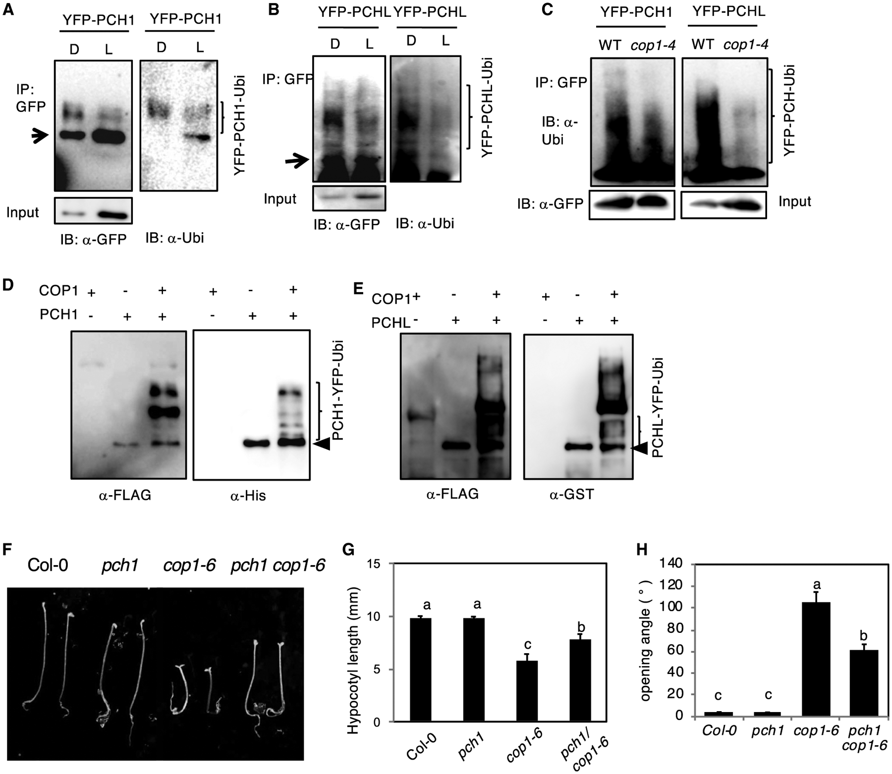
Figure 6. COP1-Mediated PCH1/PCHL Degradation Is 26S Proteasome-Dependent.

(A and B) Immunoblots show the ubiquitinated PCH1 (A) and PCHL (B) level under both dark and light.
(C) Immunoblots showing the relative ubiquitination status of YFP-PCH1 (left) and YFP-PCHL (right) in response to dark in cop1–4 mutants compared with the wild type. Total proteins were extracted from 4-day-old dark-grown seedlings and then immunoprecipitated with anti-GFP antibody (rabbit). The immunoprecipitated samples were then separated on 6.5% SDS–PAGE gels and probed with anti-GFP (mouse, left) or anti-Ubi antibodies (right). The upper smear bands are polyubiquitinated PCH1 and PCHL. Arrows indicates YFP-PCH1 and YFP-PCHL. Experiments were done three times with similar results, and a representative blot is shown.
(D and E) COP1 promotes ubiquitination of PCH1 and PCHL in vitro. An in vitro ubiquitination assay was performed for both PCH1 (D) and PCHL (E). FLAG-tagged ubiquitin was used. Left: immunoblot detected by anti-FLAG antibody. Right: immunoblot using anti-His for PCH1 and GST antibody for PCHL. Arrows indicate His-PCH1 and GST-PCHL.
(F) pch1 suppresses cop1–6 phenotype in darkness. Seedling morphology of seedlings grown under continuous darkness for 3 days. Scale bar, 5 mm.
(G and H) Bar graphs show the quantification of hypocotyl length (G) and the cotyledon opening angle (H) in (F). Error bars represent SD (n > 30 seedlings). Statistical significance among different genotypes was determined using one-way analysis of variance and Tukey’s honest significant difference tests, and is indicated by different letters.
