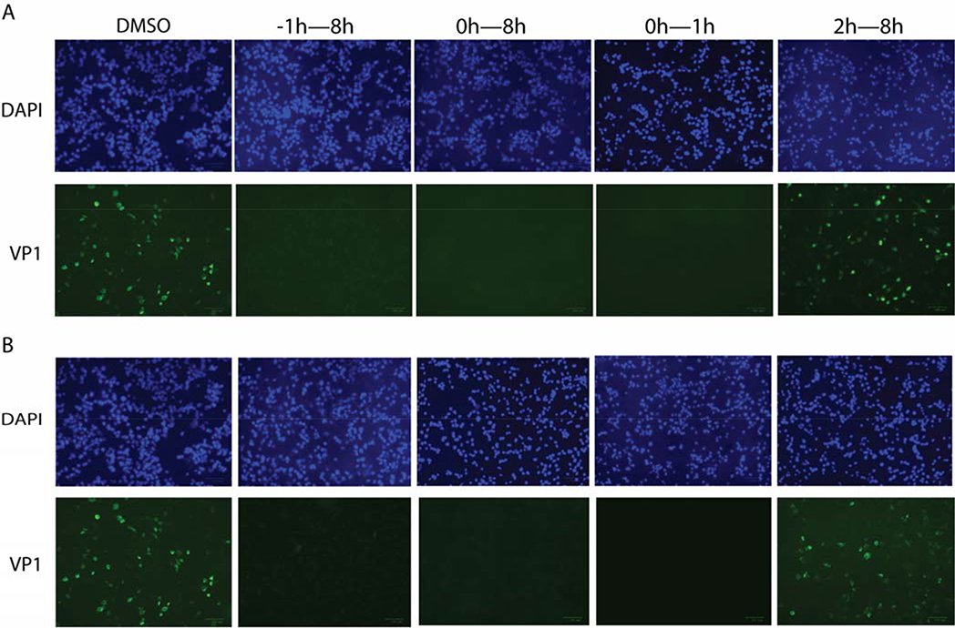
Figure 4: Time-of-addition experiments by immunostaining VP1 protein inside the host cells.
RD cells were infected with EV-D68 US/MO/14–18947 at MOI = 1 at 0 h time-point, and at 1 hpi, inoculant virus was removed and washed with PBS buffer. A) Pleconaril or B) Compound R856932 was applied at −1, 0 or 2 hpi to 8 hpi; for the 0h–1h condition, Pleconaril or compound R856932 was only present during the infection stage for one hour, then pleconaril or compound R856932 was washed away and fresh media was applied. The concentrations used for R856932 and pleconaril were 50 μM and 12.5 μM, respectively. Cells were fixed at 8 hpi and viruses were detected via staining VP1 protein with anti-VP1. The images were representatives of three independent experiments.

