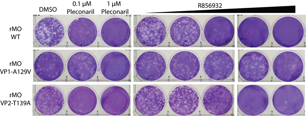
Figure 8. Representative plaque reduction assay with compound R856932 against recombinant rMO WT, rMO VP1-A129V and rMO VP2-T139A viruses.
Approximately 100 pfu/well of recombinant rMO WT, rMO VP1-A129V or rMO VP2-T139A viruses were applied to RD cell monolayers. 0.1 to 10 μM compound R856932, or 0.1 or 1.0 μM pleconaril was present in the 1.2% avicel overlay. RD cells were stained with crystal violet 3 days after infection. The images were representatives of three independent experiments.

