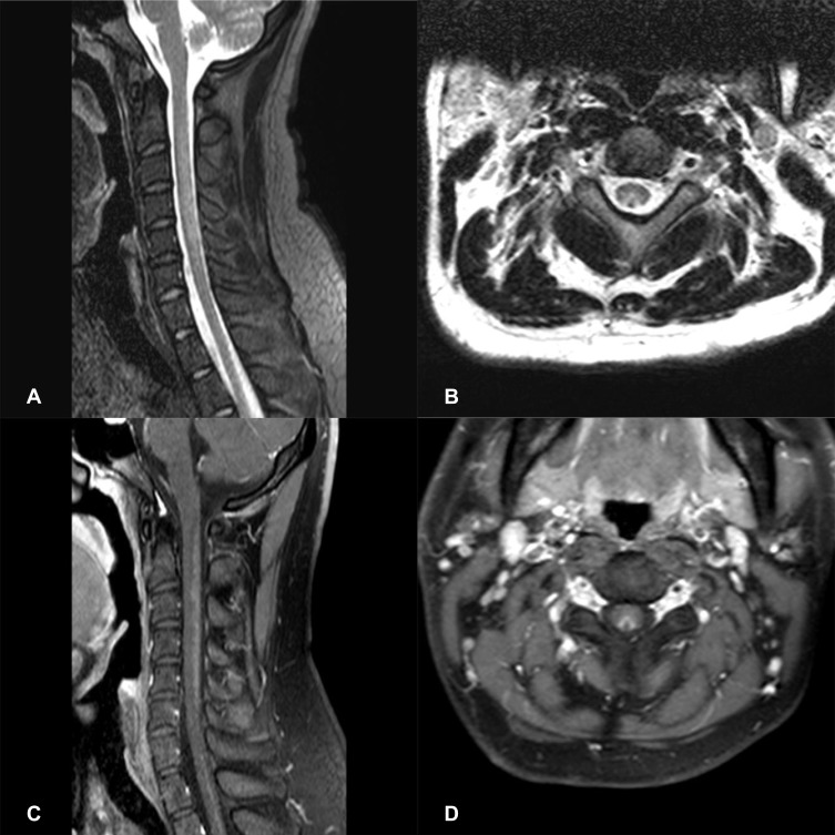
Figure 3.
Spinal cord MRI of a N2O-abuse patient who complained of unsteadiness while walking and limb numbness for 2 weeks. (A) Sagittal T2 MRI of the cervical spine demonstrating a high-signal lesion representing demyelination C2-C6. (B) Axial T2 MRI of the cervical spine shows hyperintense, symmetric and inverted V-shaped signals involving the dorsal columns of C2-C6. (C) Gadolinium-enhanced T1 MRI reveals the enhancement of the dorsal columns. (D) Gadolinium-enhanced axial T1 MRI shows a V-shaped enhancement of the dorsal columns.

