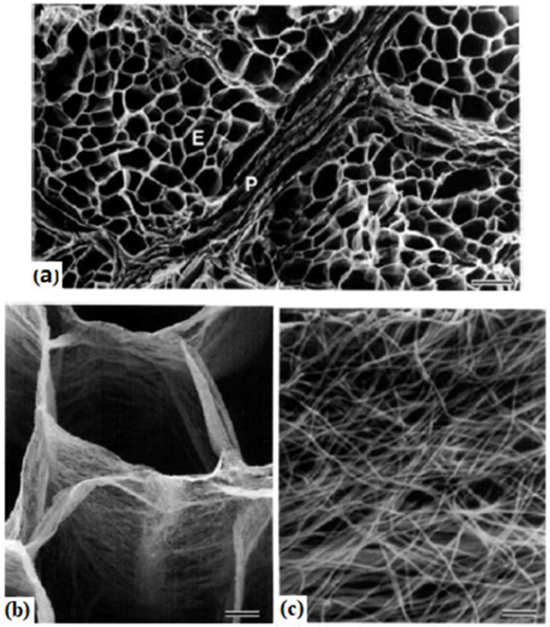
Figure 5.
Scanning electron micrographs of intramuscular connective tissue of treated bovine semitendinosus muscle using the cell-maceration method. (a) The endomysium, E, is a cylindrical sheath housing individual muscle fiber, and the perimysium, P, is composed of several layers of collagen sheets.
Bar=100μm. (b) Endomysial sheaths are membranous. Bar=10 μm. (c) A closer view of endomysium shows the random arrangement of fibrils (possibly collagen). Bar=500 nm. The figure is reproduced with permission [29]

