



Abstract
UDP-glucuronosyltransferase 2B17 (UGT2B17) is a highly variable androgen- and drug-metabolizing enzyme. UGT2B17 exhibits a unique ontogeny profile characterized by a dramatic increase in hepatic protein expression from pre-pubertal age to adulthood. Age, sex, copy number variation (CNV), and single nucleotide polymorphisms only explain 26% of variability in protein expression, highlighting the need for a phenotypic biomarker for predicting inter-individual variability in glucuronidation of UGT2B17 substrates. Here we propose testosterone glucuronide (TG) normalized by androsterone glucuronide (TG/AG) as a urinary UGT2B17 biomarker, and examine the associations between urinary TG/AG and age, sex, and CNV. We performed targeted metabolomics of 12 androgen conjugates with LC-MS/MS in 63 pediatric subjects ages 7 to 18 years followed over 7 visits in 3 years. Consistent with the reported developmental trajectory of UGT2B17 protein expression, urinary TG/AG is significantly associated with age, sex, and CNV. In conclusion, TG/AG shows promise as a phenotypic urinary UGT2B17 biomarker.
Keywords: UGT2B17, copy number variation, biomarker, testosterone glucuronide, ontogeny, interindividual variability
INTRODUCTION
UDP-glucuronosyltransferase 2B17 (UGT2B17) is a highly variable androgen- and drug-metabolizing enzyme, showing over a 3000-fold variability in hepatic protein expression.1 It is one of the most commonly deleted genes in the human genome with the frequency of the gene deletion event ranging from 16% in Caucasians up to 96% in Asians.2 However, even among expressers carrying two copies of the UGT2B17 gene, greater than 160-fold variability in expression has been reported,3 suggesting that copy number variation (CNV) is not the only determinant of the variability. Indeed, factors such as age, sex, and single nucleotide polymorphisms (SNPs) also affect UGT2B17 expression and activity. Still, only 26% of the variability can be explained by all these factors combined.4
UGT2B17 also exhibits a unique developmental trajectory. UGT2B17 is minimally expressed in children under 9 years of age (0.11 ± 0.02 pmol/mg protein) but increases approximately 10-fold throughout adolescence to adulthood (1.31 ± 0.15 pmol/mg protein) in its hepatic protein expression. The increase is much more prominent in males compared to females, and adult males have on average 2.6-fold higher levels compared to females (1.75 ± 0.20 vs. 0.65 ± 0.18 pmol/mg protein, respectively).5 Major endogenous substrates for UGT2B17 are C19 androgen steroids, such as testosterone, androsterone, and etiocholanolone.6,7 UGT2B17, when present, drives testosterone glucuronide (TG) formation, leading to highly variable TG formation.3 This has been demonstrated in vitro with recombinant enzymes, human liver microsomes, human hepatocytes and enterocytes.3,8 In contrast, UGT2B15, which is the minor isoform for TG formation with 10-fold lower activity, shows no CNV, much lower protein variability, and no age-dependency.1,8,9
High variability in UGT2B17 expression has shown in vivo associations with several important outcomes. UGT2B17 gene deletion is associated with increased risk of prostate cancer, amidst conflicting results.10 Other testosterone-related outcomes, such as lower body mass index (BMI)11 and increased bone mineral density12 are associated with UGT2B17 deletion. However, other studies report conflicting results,13 possibly due to the high interindividual variability in UGT2B17-mediated testosterone metabolism. In addition, UGT2B17 genotype is a well-known confounder in antidoping tests for athletes, where the urinary testosterone to epitestosterone ratio (T/E ratio) is measured after deconjugation with β-glucuronidase.14
UGT2B17 variability has led to unexpected pharmacokinetic (PK) outcomes resulting in drug development failures. For example, an antagonist of chemoattractant receptor-homologous molecule expressed on T-helper type 2 cells (CRTH2), MK-7246, failed its first-in-human study due to unexpected PK variability, and UGT2B17 was retrospectively identified as the primary source of variability, with 25-fold higher systemic exposure in subjects with two gene copies compared to those homozygous for a gene deletion event.15 Similarly, unpredictable high variability in the metabolism of PT2385, an inhibitor of hypoxia-inducible factor-2α, led to discontinuation of its development program.16 UGT2B17 variability also affects drug disposition in currently utilized therapeutics. 17-hydroexemestane, the major active metabolite of the oral antineoplastic drug exemestane, showed significantly higher exposure in patients homozygous for UGT2B17 deletion compared to those with 2 copies, presumably due to reduced glucuronidation.17 Additionally, metabolism of the oral antineoplastic agent vorinostat is significantly affected by UGT2B17 CNV, which is reflected by trends in higher adverse events and longer median progression-free survival for patients with no UGT2B17 copies compared to those with at least 1 copy of the gene.18
Despite these in vivo reports, UGT2B17 receives limited attention from regulatory agencies and pharmaceutical industries compared to other UGTs, possibly due to the small number of clinically approved compounds primarily metabolized by UGT2B17, along with its minor presence in commonly used in vitro models such as pooled liver microsomes. However, UGT2B17 can play a significant role in drug metabolism and drug-drug interactions (DDIs) in high UGT2B17 expressers (e.g., individuals with 100-fold higher abundance compared to low expressers). Furthermore, intestinal UGT2B17 expression is on average 4-fold higher than liver,3 and UGT2B17 is the major UGT isoform expressed in the intestine, with a 3- and 7-fold higher expression than UGT1A1 and UGT2B7, respectively.1 High and variable intestinal expression of UGT2B17 may significantly affect drug bioavailability.15 Variable effects also have potential to be compounded in children due to steep increase in UGT2B17 expression during development, especially for chronic medications, where dose adjustment may be warranted to address the rapidly changing UGT2B17 expression.
An endogenous phenotypic biomarker may be of considerable value to predict UGT2B17 activity in vivo, considering the potential role of UGT2B17 in xenobiotic and endobiotic disposition, association with pathophysiological outcomes, and high interindividual and age-related variability. Specific and sensitive biomarkers offer advantages in drug development by reducing unnecessary and costly DDI trials and avoiding late-stage discontinuations,19 and improves patient safety by its less invasive nature, as recognized by United States Food and Drug Administration (FDA).20
Urinary TG has shown strong association with UGT2B17 activity in children and adults,21,22 but TG alone as a biomarker is confounded by differing levels of endogenous testosterone synthesis. Although testosterone (T) would be the ideal normalizer, it is undetectable in urine. Thus, we propose androsterone glucuronide (AG) as a normalizer for TG. AG normalization should control for the interindividual variability in endogenous testosterone production. Androsterone is an inactive androgen, is reported to represent peripheral androgen metabolism, and its conjugated metabolite AG is abundant in urine and blood.23 AG formation is primarily mediated by UGT2B7, which has substantially less variability and relatively unchanging ontogeny during pubertal ages,1,9 with negligible contribution from UGT2B17 (Figure 1).8 Total (conjugated and deconjugated) urinary epitestosterone is a common normalizer used in T/E ratio for anti-doping tests; however, this aims to differentiate exogenous testosterone administration. Epitestosterone does not address the fluctuations in endogenous testosterone production, as it is regulated by a separate pathway.23 Epitestosterone also exhibits poorer sensitivity due to lower urinary levels and is susceptible to effects of other polymorphisms, menstrual cycles, and drugs.24,25 Polymorphisms, allosteric regulations, reversibility, and CNV of sulfotransferases limit the potential for normalization with sulfate conjugates.26
Figure 1. Androgen metabolism scheme.
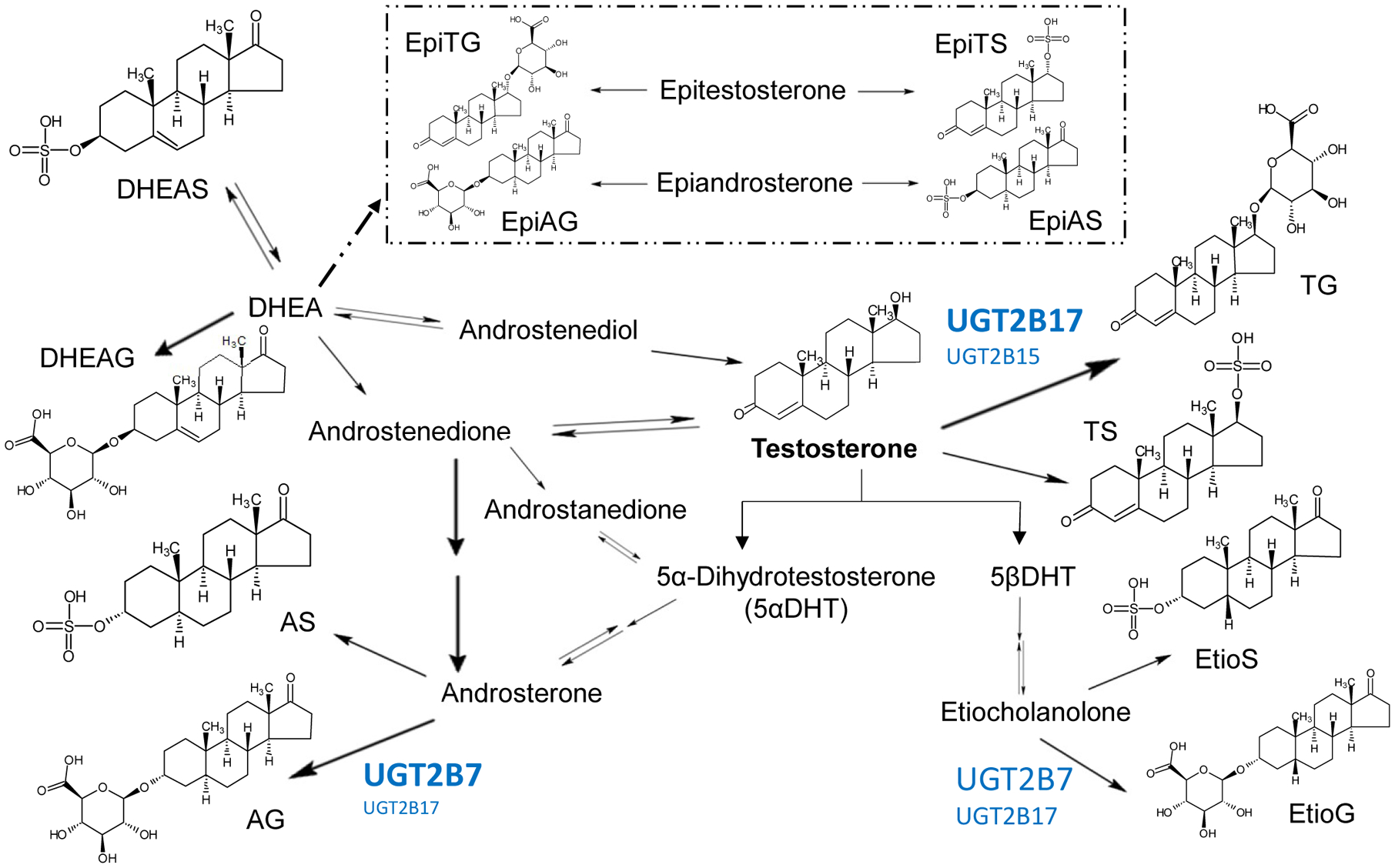
Simplified androgen metabolism scheme49,50 is shown, with relative UGT contribution in glucuronidation depicted in blue. Dashed arrow and box indicate a separately regulated metabolic pathway for epitestosterone and epiandrosterone that is not fully understood.
In this study, we investigated TG normalized by androsterone glucuronide (TG/AG) as a potential noninvasive urinary biomarker for UGT2B17 activity by its association with age, sex, and CNV state using longitudinal pediatric urine samples and targeted metabolomics by LC-MS/MS. Targeted analyses of ten other androgen conjugates were done to examine associations with age, sex, and CNV state. In addition, we performed exploratory analysis to investigate associations of race and sexual maturity rating (SMR) with urinary TG/AG levels.
METHODS
Clinical study details and sample collection
Pediatric urine samples were used from a previous longitudinal phenotyping study conducted at Children’s Mercy Hospital, Kansas City, MO, USA.27 Samples were collected in seven visits over a three-year period, with subject ages ranging from 7 to 18 years old. Analyzed urine samples were obtained in the morning prior to dosing and were previously unthawed from −80°C storage.
Urine sample processing
Solid phase extraction (SPE) was performed using Oasis® hydrophilic-lipophilic balanced (HLB) 1 mL cartridges with 30 mg sorbent (Waters Technologies, Milford, MA). 300 μL of distilled water containing internal standards (IS) was added to 300 μL aliquot of urine and vortexed. HLB cartridges were conditioned with methanol and equilibrated with distilled water. Samples were loaded and passed through, followed by wash with 2% formic acid in 5% methanol in water. Elution was collected with 5% ammonia in methanol, evaporated under nitrogen stream, and reconstituted with 100 μL of 1:1 methanol: water. Processed samples were stored in −80°C prior to LC-MS/MS analysis. Quality controls and pooled samples were included in each batch processing, to control for technical and instrumental variabilities.
LC-MS/MS method development, validation and analysis of urine androgen conjugates
Glucuronide (G) and sulfate (S) conjugates for 6 androgens were analyzed: testosterone (T), androsterone (A), etiocholanolone (Etio), epitestosterone (EpiT), epiandrosterone (EpiA), and dehydroepiandrosterone (DHEA). LC-MS/MS analysis was performed using Sciex Triple Quadrupole 6500 system (Sciex, Framingham, MA) coupled with Acquity UPLC System (Waters Technologies, Milford, MA). ACQUITY UPLC HSS T3 1.8 μm, C18 100A; 100 × 2.1 mm (Waters Technologies, Milford, MA) column was used to achieve chromatographic separation of conjugates (Supplementary Figure S1). LC conditions had a flow rate of 0.3 mL/min with 5 μL injection volume. Mobile phase A1 and A2 were 0.1% formic acid in water and 10 mM ammonium acetate in water with 0.1% acetic acid and B1 and B2 were 0.1% formic acid in acetonitrile and 0.1% acetic acid in acetonitrile. A1 and B1 were used for glucuronides and A2 and B2 for sulfates. Gradient programs were set as follows: 75% A1 (0–1 min), 75–60% (1–6 min), 60–58% (8–9 min), 58 to 50% (9–10 min), 50–10% (10–12 min), 10–75% (13–13.1 min), 75% (13.1–15 min) for glucuronides, and 74% A2 (0–1 min), 74–71% (1–6 min), 71–63% (6–12 min), 63–5% (12–12.5 min), 5–73% (13–13.1 min), 73% (13.1–15 min) for sulfates. Positive and negative ESI mode was used for glucuronides and sulfates, respectively, and MS parameters used for quantification are shown in Table 4. Acquired data was processed using Skyline daily software (University of Washington, Seattle, WA). Lower limit of quantifications (LLOQ) were established, and the lower limit of detection (LOD) was set as one-third of LLOQ (Table 4). Samples that were below limit of quantification (BLOQ) were assigned LOD values and included for analysis. US FDA guidance for bioanalytical method validation (https://www.fda.gov/files/drugs/published/Bioanalytical-Method-Validation-Guidance-for-Industry.pdf) was followed for method validation.
Table 4.
MS parameters for targeted androgen conjugates
| Metabolite | Precursor ion (m/z) | Product Ion(s) (m/z) | DP (V) | CE (eV) | LLOQ# (ng/mL) |
|---|---|---|---|---|---|
| GLUCURONIDES | |||||
| Androsterone glucuronide (AG) | 449.1 | 255 | 70 | 25 | 2 |
| Androsterone glucuronide-d4* | 453.1 | 259 | 70 | 25 | - |
| Etiocholanolone glucuronide (EtioG) | 484.3 | 273, 255 | 70 | 35 | 1 |
| Testosterone glucuronide (TG) | 465.2 | 289, 271, 253, 109, 97 | 70 | 25, 30, 35, 40, 35 | 0.5 |
| Testosterone glucuronide-d3* | 468.2 | 292, 274, 256, 109, 97 | 70 | 25, 30, 35, 40, 35 | - |
| DHEA+ glucuronide (DHEAG) | 465.2 | 289, 271, 253, 109, 97 | 70 | 25, 30, 35, 35, 35 | 25 |
| Epitestosterone glucuronide (EpiTG) | 482.2 | 253, 271, 252 | 50 | 25, 30, 37 | 1.5 |
| Epiandrosterone glucuronide (EpiAG) | 449.1 | 273, 255 | 70 | 25, 30 | 7.5 |
| SULFATES | |||||
| Androsterone sulfate (AS) | 369.2 | 97, 80 | −155 | −78, −130 | 2.5 |
| Androsterone sulfate-d4* | 373.2 | 97, 80 | −155 | −78, −130 | - |
| Etiocholanolone sulfate (EtioS) | 369.2 | 97, 80 | −150 | −90, −118 | 5 |
| Etiocholanolone sulfate-d5* | 374.2 | 97, 80 | −150 | −90, −118 | - |
| Testosterone sulfate (TS) | 367.2 | 351, 97, 80 | −165 | −54, −84, −120 | 2.5 |
| Testosterone sulfate-d3* | 370.2 | 354, 97, 80 | −165 | −54, −84, −120 | - |
| DHEA+ sulfate (DHEAS) | 367.2 | 97, 80 | −120 | −72, −114 | 3 |
| DHEA+ sulfate-d5* | 372.2 | 97, 80 | −120 | −72, −114 | - |
| Epitestosterone sulfate (EpiTS) | 367.2 | 97, 80 | −140 | −80, −124 | 0.5 |
| Epitestosterone sulfate-d3* | 370.2 | 97, 80 | −140 | −80, −124 | - |
| Epiandrosterone sulfate (EpiAS) | 369.2 | 97, 80 | −125 | −88, −116 | 3 |
deuterated internal standard,
dehydroepiandrosterone,
lower limit of quantification
UGT2B17 gene copy number determination
CNV analysis was performed at Children’s Mercy Hospital (Kansas City, MO) and are described elsewhere in detail.28 In brief, quantitative multiplex PCR amplification (MPA) was performed, fragments separated on an ABI 3730 instrument, and subsequently analyzed with GeneMapper v5 (both Thermo Fischer, Waltham, MA). UGT2B17 copy number was determined by normalizing against UGT2B15 which is not known to undergo copy number variation.
Statistical analysis
Statistical analysis was performed using GraphPad Prism 5.03 (La Jolla, CA) and R (version 3.5.1). Linear correlation tests between CNV and subject demographic variables were performed using data from the first visit. Similarly, correlations of age, sex, and CNV with other androgen conjugates were examined.
TG/AG was log-transformed for normality. To assess the association of log (TG/AG) with age and sex, we used a linear spline model with a generalized estimating equation for population inference, after exclusion of CNV=0 subjects (Equation 1). Age splines were set at 11 and 14 years, to incorporate the average male onset ages of SMR stages 2 and 4, respectively. These age knots were selected to enable continuous assessment of changes in TG/AG and to account for disproportionate increases in reported urinary androgens between SMR 2 and 4.29 Age spline settings were also consistent with exploratory LOWESS curve fitting.
| Equation 1: |
The β coefficient indicates the rate of change in log (TG/AG) for the following variable, and i is the index for each subject, while j is the index for each time point. The + sign sets the lower boundary as zero, allowing age spline formation based on the distance to the knots (e.g. 11 and 14 years).
We examined the association of log (TG/AG) with CNV and age separately in males and females, after observation of significant trend differences in sex and age. Linear spline model was applied, and CNV was treated as a continuous variable to incorporate increasing trend of TG/AG with increasing copy number (Equation 2).
| Equation 2: |
The association of log (TG/AG) with race (i.e., African American and Caucasian) was explored using equation 3:
| Equation 3: |
Given the physiological as well as statistical correlation between age and SMR, we also looked at association with SMR (Equation 4), with splines set at stages 2 and 4, reflecting the average age splines from Equation 2.
| Equation 4: |
Fitted model outputs for β coefficients were tested against the null hypothesis of β =0 (no association) and considered significant at p <0.05.
RESULTS
Bioanalytical method validation
We developed a sensitive and specific LC-MS/MS-based method to analyze both glucuronide (G) and sulfate (S) conjugates for six androgens: testosterone (T), epitestosterone (EpiT), androsterone (A), epiandrosterone (EpiA), etiocholanolone (Etio), and dehydroepiandrosterone (DHEA). Sensitivity was established with LLOQ and specificity was confirmed, and accuracy and precision across different runs were within 20% for both for TG and around 30% and 15%, for AG, respectively. Different measures for variability and stability testing were performed, including interday variability, intraday variability, and interanalyst variability, as well as freeze-thaw stability, benchtop stability, 1-year long-term stability, and autosampler stability. Interday, intraday and interperson variability (%CV) was 14.1% and 6.9% for TG, and 16.4% and 7.8% for AG, respectively. Freeze-thaw, benchtop, long-term and autosampler stability (% difference) was 1.3%, 3.4%, 8.9%, and 0.4% for TG, and 7.7%, 11.7%, 4.6%, and 4.1% for AG, respectively. Most other conjugates were acceptable within 20%; EtioG was readily detected but not quantifiable due to IS normalization issues. Recovery for TG was 111% and AG was 55%.
Urinary concentrations of androgen conjugates
Androgen conjugates were quantified in urine samples (n=439) collected for 63 subjects from 7 visits over 3 years (Table 1). Average urinary concentrations and standard deviation for quantified androgen conjugates in male and female age groups (<11, 11–14, and >14 years) are shown in Table 2, with DHEAG and EpiAG mostly below the limit of quantification, and EtioG not quantified as explained above. TG and EpiTG concentrations increased with age, and the percentage of samples with TG and EpiTG concentrations below the lower limit of quantitation decreased from 29–48% and 71–94% for males and females under 11 years of age, respectively, to 0–17% and 4–9% in the >14 years age group. T and EpiT sulfate levels were comparatively much lower than glucuronides—TS was undetectable in 50% and 89% of samples, compared to 20% and 21% for TG, for male and females, respectively.
Table 1.
Subject demographics
| Overall1 | CNV=0 | CNV=1 | CNV=2 | CNV correlation | |||||
|---|---|---|---|---|---|---|---|---|---|
| Male | Female | Male | Female | Male | Female | p value | |||
| Sex (%) | 39 M (62) 24 F (38) | 6 (10) | 4 (6) | 17 (27) | 9 (14) | 16 (25) | 11 (17) | 0.85 | |
| Age (years) | 12 ± 2.5 | 11.8 ± 2.9 | 11.6 ± 3.0 | 12.3 ±2.1 | 12.1 ± 2.2 | 12.6 ± 2.2 | 11.4 ± 2.2 | 0.72 | |
| BMI2 (kg/m2) | 20.8 ± 4.8 | 20.7 ± 3.1 | 19.8 ± 0.9 | 19.6 ±3.6 | 23.8 ± 7.2 | 21.2 ± 6.3 | 22.9 ± 5.0 | 0.93 | |
| Ethnicity3 (%) | AA | 28 (44) | 1 (2) | 5 (8) | 5 (8) | 10 (16) | 7 (11) | ref | |
| CA | 28 (44) | 5 (8) | 4 (6) | 9 (14) | 2 (3) | 4 (6) | 4 (6) | 0.0015** | |
| AA/NH | 1 (2) | 1 (2) | 0.41 | ||||||
| CA/AA | 4 (6) | 2 (3) | 2 (3) | 0.84 | |||||
| CA/NH | 2 (3) | 2 (3) | 0.25 | ||||||
| SMR4 (%) | 1 | 125 (28) | 14 (3) | 7 (2) | 49 (11) | 10 (2) | 28 (6) | 17 (4) | ref |
| 2 | 87 (20) | 10 (2) | 7 (2) | 24 (5) | 14 (3) | 23 (5) | 9 (2) | 0.68 | |
| 3 | 61 (14) | 4 (1) | 2 (0.5) | 10 (2) | 8 (2) | 21 (5) | 16 (4) | 0.37 | |
| 4 | 79 (18) | 6 (1) | 6 (1) | 18 (4) | 11 (3) | 18 (4) | 20 (5) | 0.48 | |
| 5 | 87 (20) | 8 (2) | 6 (1) | 17 (4) | 20 (5) | 22 (5) | 14 (3) | 0.69 | |
63 subjects were followed over 7 visits with total 439 urine samples analyzed
BMI: Body mass index;
CA: Caucasian American, AA: African American, NH: Non-Hispanic;
SMR: Sexual maturity rating
Table 2.
Urinary concentrations (ng/mL) of androgen conjugates in children age 7–18 years
| < 11 years | 11–14 years | >14 years | ||||
|---|---|---|---|---|---|---|
| Male | Female | Male | Female | Male | Female | |
| (n=85) | (n=70) | (n=113) | (n=62) | (n=74) | (n=35) | |
| GLUCURONIDES1 | ||||||
| TG | 1.8 ± 2.2 (48) | 3.3 ± 3.8 (29) | 20 ± 31 (12) | 6.6 ± 5.7 (15) | 75 ± 69 (0) | 9.8 ± 9.7 (17) |
| AG | 1216 ± 894 | 1273 ± 883 | 3016 ± 2306 | 3450 ± 2281 | 6826 ± 3468 | 4924 ± 2482 |
| EtioG2 | Detected2 | |||||
| DHEAG3 | BLOQ3 | |||||
| EpiTG | 0.4 ± 1.4 (94) | 1.2 ± 2.1 (71) | 5.5 ± 9.6 (44) | 5.1 ± 4.8 (11) | 22 ± 25 (4) | 9.7 ± 8.1 (9) |
| SULFATES1 | ||||||
| TS | 0.32 ± 0.27 (87) | 0.27 ± 0.11 (96) | 0.89 ± 1.81 (51) | 0.36 ± 0.33 (82) | 4.18 ± 9.41(7) | 0.34 ± 0.27 (89) |
| AS | 296 ± 327 | 156 ± 164 | 461 ± 439 | 356 ± 502 | 613 ± 497 | 534 ± 451 |
| EtioS | 106 ± 104 | 93 ± 85 | 112 ± 124 | 190 ± 174 | 114 ± 119 | 282 ± 263 |
| DHEAS | 199 ± 361 | 67 ± 109 | 430 ± 851 | 550 ± 2307 | 694 ± 1402 | 1668 ± 4154 |
| EpiTS | 2.9 ± 1.8 (5) | 2.0 ± 1.5 (10) | 6.4 ± 5.3 (2) | 3.4 ± 2.8 (13) | 18 ± 12 (0) | 4.7 ± 3.4 (3) |
| EpiAS | 44 ± 59 | 17 ± 19 | 73 ± 89 | 55 ± 111 | 98 ± 95 | 97 ± 104 |
mean ± SD (%BLOQ, otherwise all detected)
Absolute quantification not performed due to problems with normalization
Below limit of quantification (BLOQ) were assigned lower limit of detection (LOD) calculated as LLOQ/3
Association of urinary androgen conjugates with sex, age, and UGT2B17 copy number variation (CNV)
Average age for males (n=39, 62%) were 12.4 years and for females (n=24, 38%) were 11.7 years. CNV distribution were as follows (n, average age in years): for males, CNV0 (n=6, 11.8 years), CNV1 (n=17, 12.3 years), CNV2 (n=16, 12.6 years), and for females, CNV0 (n=4, 11.6 years), CNV1 (n=9, 12.1 years), CNV2 (n=11, 11.4 years) (Table 1). CNV showed no association with sex, age, body mass index (BMI), or sexual maturity rating (SMR), but was significantly associated with race, i.e. Caucasian Americans (CA) and African Americans (AA) with AA as the reference group (p=0.0015). Simple correlation analyses showed significant associations with age but not with sex for most metabolites. Androgen metabolites except EtioS (p=0.0405) showed no significant associations with UGT2B17 CNV (data not shown).
TG, AG, and TG/AG in males and females with age and by CNV is shown in Figure 2. Age-related increase in TG was more prominent in males compared to females, while increase in AG was similar in both sexes (Figure 2A and 2B), and CNV association is seen only with TG (Figure 2D and 2E). TG/AG normalization resulted in a more distinct spread (Figure 2C), and a visual association with CNV is still present for different SMR groups, which reduces for the influence of sexual maturation (Figure 2F and 2G).
Figure 2. TG, AG, and TG/AG with age and CNV, by sex.
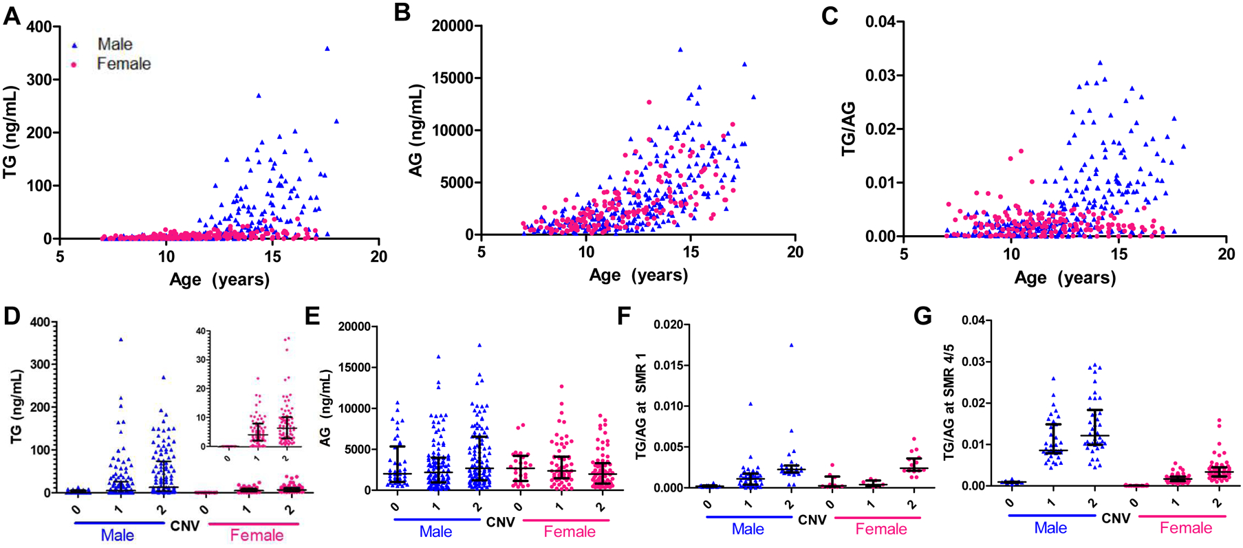
Testosterone glucuronide (TG), androsterone glucuronide (AG), and TG/AG with age is shown in A, B, and C, and TG and AG with copy number variation (CNV) is shown in D and E, respectively. TG/AG with CNV is separated by sexual maturity rating (SMR) at stage 1 (F) and stages 4 and 5 (G), to remove the influence of sexual maturation. Males are depicted in blue triangles and females in pink circles; line and bars show median and interquartile ranges.
TG/AG associations with age, sex, race, and CNV were all significant (p<0.001) (Table 3). Fitted linear spline model for log (TG/AG) with age for male and female is shown in Figure 3A. Figure 3B shows the fitted model by race (CA and AA). The significant difference between the two races in this analysis is likely due to different CNV frequencies in CA and AA as a confounder. Earlier correlation analysis showed significant association between CNV and race (Table 1), thus genetic differences in CNV frequency may manifest as racial differences. It has been reported that the UGT2B17 deletion allele is five times more frequent in Caucasians (11%) than African Americans (2%).30 Fitted models for log (TG/AG) by CNV with age in male and females are shown in Figures 3C and 3D, respectively. Log (TG/AG) was also associated with SMR (p<0.001).
Table 3.
Linear regression outputs of β coefficient values
| Age and sex | Age and CNV (Male) | Age and CNV (Female) | Age and Race | CNV and SMR (Male) | CNV and SMR (Female) | Significance# | |
|---|---|---|---|---|---|---|---|
| β0 | −6.69 | −8.36 | −9.96 | −6.21 | −8.32 | −8.71 | *** |
| β1 | 1.25 | 1.07 | 1.65 | −2.41 | 0.92 | 0.93 | *** |
| β2 | 0.06 | −0.04 | 0.13 | −0.01 | −0.18 | 0.05 | *** |
| β3 | −0.12 | 0.78 | −0.33 | 0.33 | 1.41 | −0.41 | *** |
| β4 | 0.00 | −0.61 | 0.08 | −0.34 | −1.26 | 0.62 | *** |
| β5 | −0.20 | 0.03 | −0.01 | 0.15 | 0.32 | 0.43 | *** |
| β6 | 1.06 | −0.10 | 0.06 | −0.14 | −0.59 | −0.38 | *** |
| β7 | −0.73 | 0.05 | 0.08 | 0.15 | 0.33 | −0.08 | *** |
Statistically significant for all β coefficient values for H0: β=0
Figure 3. Association of urinary TG/AG levels with age, sex, race, and CNV.
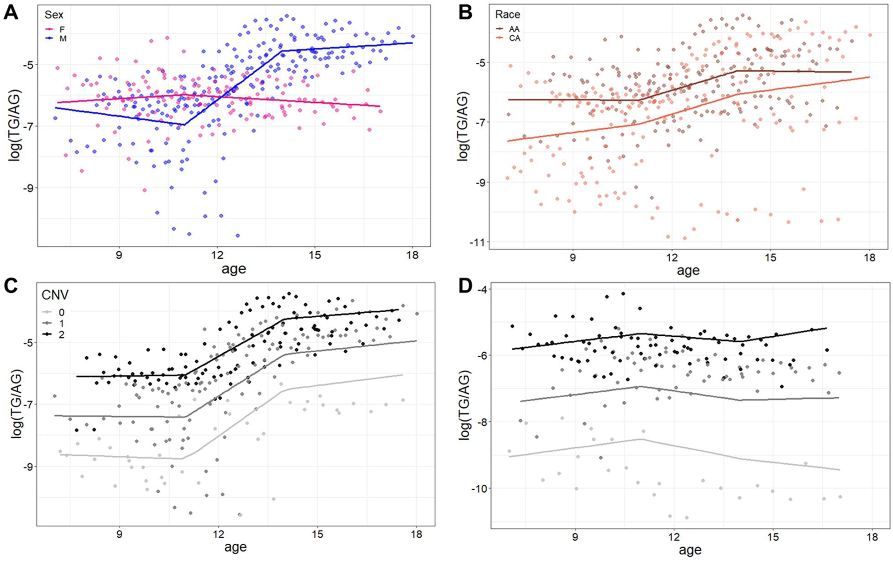
Model-fitted log (TG/AG) with age splines set at 11 and 14 years is shown with males in blue and females in pink (A). Log (TG/AG) association with race is shown in B, with African Americans (AA) in dark brown and Caucasian Americans (CA) in light brown. Log (TG/AG) association with copy number variation (CNV=0 in light grey, CNV=1 in dark grey, CNV=2 in black) is shown for males (C) and females (D).
DISCUSSION
We quantified a panel of androgen conjugates in longitudinal pediatric urine samples, with a comprehensive analysis of association between TG/AG with multiple variables including genotype and SMR. Urinary concentrations of androgen conjugates were in agreement with available reported ranges.22,23,29 The observed increase in urinary TG with UGT2B17 CNV has also been reported, and is supported by the greater than 20-fold difference seen between CNV=2 and CNV=0.21 EtioS association with UGT2B17 CNV that was observed has also previously been reported.31 While we showed TG/AG showed significant association with SMR, urinary testosterone to epitestosterone ratio (T/E) is reported to be invariant between SMR stages.29 Consistent with our hypothesis, TG/AG showed significant associations with age, sex, and CNV state, with a steeper increase in males compared to females, which also visually matches UGT2B17 ontogeny profile in hepatic protein abundance from a separate dataset.5
Once validated as a UGT2B17 biomarker, TG/AG has potential utilization in better prediction of drug disposition, DDIs, interindividual variability, and study design in children and adults. For example, urinary TG/AG levels of subjects in Phase I clinical trials could predict if an investigational drug is a likely inhibitor of UGT2B17. This application is important as UGT2B17 inhibition is not routinely tested during early drug development. TG/AG may also be used for in vivo predictions of the variable activity and contribution in drug disposition. Acyl glucuronide metabolites such as gemfibrozil glucuronide and clopidogrel acyl glucuronide, (CAG) have recently gained attention as potent cytochrome P450 2C8 inhibitors that lead to clinically significant DDIs.32 In particular, UGT2B17’s contribution in CAG formation is estimated to be on average around 10% and UGT2B7 as a major contributor.33 However, in high UGT2B17 expressers, CAG formation and subsequent DDI can significantly increase, but potential DDI magnitudes may be screened using TG/AG. Stratification of study subjects as null, mid, and high UGT2B17 expressers rather than CNV=0, CNV=1, CNV=2, may also aid in more robust and reliable results.
Given UGT2B17’s role in androgen metabolism, TG/AG biomarker may also elucidate the role of UGT2B17 in pubertal development. The UGT2B17 deletion allele is associated with delayed puberty in 668 healthy boys, with 0.34 years delay in pubarche onset per deletion allele, but no differences in circulating testosterone or DHEAS.34 This suggests that UGT2B17 may play an important part in androgen intracrinology, where regulation of androgen activation and metabolism occur within the cell, with minimal release of active androgens into systemic blood circulation. UGT2B17 may also play a part in disease susceptibility, such as development of polycystic ovary syndrome (PCOS), where ethnic differences have also been observed.35 TG/AG may also aid in controlling for UGT2B17 confounding for better robustness of antidoping tests.
The association between the UGT2B17 deletion allele and cancer is very intriguing due to bidirectional associations with cancer risk versus cancer progression. Absence of UGT2B17 gene (CNV=0) seems to increase the risk of cancer for prostate,10 breast, pancreatic,36 and lung37 cancer, but decrease the risk of colorectal cancer.38 On the other hand, overexpression of UGT2B17 protein has been linked to biochemical recurrence of prostate cancer,39 expedition in progression to castration-resistant prostate cancer,40 and poorer prognosis in chronic lymphocytic leukemia due to inactivation of protective prostaglandin E2.41 While presence of UGT2B17 gene may be onco-protective, its seemingly deleterious role in cancer progression suggests a separate function that is under differential regulation. However, much is still unknown regarding UGT2B17 regulation, in both normal physiology as well as abnormal proliferation. It is unknown if overexpression in tumors is related to higher baseline expression levels of UGT2B17. Conflicting findings on CNV-based associations may partly be due to the variability in UGT2B17. A biomarker (TG/AG) could potentially allow for baseline stratification based on UGT2B17 expression levels without solely relying on CNV state, possibly yielding more conclusive results.
Limitations of the study include the small sample size of 63 subjects and the relatively short follow-up period of 3 years. Both these factors may partially explain the relatively flat TG/AG trend with age seen in females, although conclusive associations could still be drawn. Study demographics had no Asians to allow further exploration of race; however, we did see ethnic differences between Caucasians and African Americans, which is likely attributable to differences in CNV frequency. Although urine samples were collected in the morning in this study, it is known that testosterone undergoes diurnal variation. However, the degree of variation for testosterone is reported to be not as dramatic compared to cortisol. Androsterone also undergoes similar fluctuations, and normalization by AG would further dilute the effect of diurnal variation.42,43 Further clinical validation studies that provide a direct UGT2B17 activity relationship, such as using an exogenous probe substrate, would be beneficial to establish TG/AG ranges for phenotypic groupings (null, low, moderate, and high) that can be used to guide clinical decisions or study design. However, the lack of UGT2B17-specific probe substrate that are proven safe for pediatric administration poses another limitation. In addition, lower levels of TG excretion in UGT2B17 deletion individuals and women poses problems in TG detectability, and instrumental sensitivity is an area for improvement. Low levels of DHEAG and EpiAG and incomplete quantification of EtioG are other limitations. There are potential limitations of TG/AG as a biomarker. TG/AG is more indicative of hepatic UGT2B17 expression, considering the sites of androgen synthesis and the relative systemic blood flow to liver versus the intestine. However, up to ten percent of TG also undergoes enterohepatic recycling,44 so systemic TG/AG also incorporates intestinal UGT2B17, although more studies are needed on co-regulation of hepatic and intestinal UGT2B17. Numerous enzymes are involved in steroidogenesis and pubertal development,23 Steroid- or steroidal enzyme- related disorders, such as precocious puberty or hyperandrogenism, may alter endogenous production and TG or AG levels, rendering TG/AG unreliable for UGT2B17 estimation. TG/AG also cannot be used when UGT2B7 expression is significantly affected, such as cirrhosis.45 Moreover, efflux transporters multidrug resistance-associated protein 2 (MRP2) and MRP3 contribute to the apical and basolateral transport of TG and AG, respectively, in the liver, intestine, and kidneys.44,46 Both transporters show low interindividual variability as well as insignificant ontogeny in kidney and liver.47,48 However, additional characterization of TG and AG disposition, including effects of transporter and enzyme polymorphisms would be beneficial for further studies, especially in UGT2B17 deletion subjects.
In conclusion, urinary TG/AG was associated with age, sex, and UGT2B17 CNV. TG/AG is a promising biomarker to characterize the highly variable UGT2B17 activity in children 7 to 18 years old. Clinical studies using UGT2B17 probe substrates are needed for validation of TG/AG as a UGT2B17 biomarker. Once validated, TG/AG holds potential applications in non-invasive in vivo assessment of UGT2B17 activity for prediction of interindividual variability, DDIs, as well as disease-state associations.
Supplementary Material
Urinary androgen conjugates: chromatographic separation and concentrations (ng/mL) by age (years) and by CNV state
Study Highlights.
What is the current knowledge on the topic?
UGT2B17 shows over a 3000-fold inter-individual variability with a common gene deletion. However, gene deletion, sex, age, and genetic polymorphisms only account for 26% of the variability in UGT2B17 expression. UGT2B17 shows a distinct ontogeny profile, with a steep increase in protein expression during adolescence, and the steepness is more prominent in males than females.
What question did this study address?
Does TG/AG show associations with age, sex, and CNV in pediatric urine samples as a potential phenotypic urinary biomarker for UGT2B17?
What does this study add to our knowledge?
TG/AG is a novel biomarker proposed to more accurately determine UGT2B17 activity or phenotype.
How might this change clinical pharmacology or translational science?
TG/AG may be useful in assessing drug interactions, designing clinical studies, investigating pubertal development, and elucidating other UGT2B17-associated diseases.
Acknowledgements:
Authors would like to thank Dr. Timothy Thorton and Dr. Kenneth Thummel for their insightful suggestions.
Funding information: This work was supported by the National Institutes of Health (NIH) grant (R01 HD058556 and R01 HD081299) and Department of Pharmaceutics, University of Washington.
Footnotes
Conflict of interest: All authors declared no competing interests for this work.
References:
- 1.Oda S, Fukami T, Yokoi T & Nakajima M A comprehensive review of UDP-glucuronosyltransferase and esterases for drug development. Drug Metab. Pharmacokinet 30, 30–51 (2015). [DOI] [PubMed] [Google Scholar]
- 2.Xue Y et al. Adaptive evolution of UGT2B17 copy-number variation. Am. J. Hum. Genet 83, 337–346 (2008). [DOI] [PMC free article] [PubMed] [Google Scholar]
- 3.Zhang H et al. Quantitative characterization of UDP-glucuronosyltransferase 2B17 in human liver and intestine and its role in testosterone first-pass metabolism. Biochem. Pharmacol 156, 32–42 (2018). [DOI] [PMC free article] [PubMed] [Google Scholar]
- 4.Bhatt DK et al. Hepatic Abundance and Activity of Androgen and Drug Metabolizing Enzyme, UGT2B17, are Associated with Genotype, Age, and Sex. Drug Metab. Dispos dmd.118.080952 (2018).doi: 10.1124/dmd.118.080952 [DOI] [PMC free article] [PubMed] [Google Scholar]
- 5.Bhatt DK et al. Hepatic Abundance and Activity of Androgen- and Drug-Metabolizing Enzyme UGT2B17 Are Associated with Genotype, Age, and Sex. Drug Metab. Dispos 46, 888–896 (2018). [DOI] [PMC free article] [PubMed] [Google Scholar]
- 6.Hum DW et al. Characterization of UDP-glucuronosyltransferases active on steroid hormones. J Steroid Biochem Mol Biol 69, 413–423 (1999). [DOI] [PubMed] [Google Scholar]
- 7.Turgeon D, Carrier JS, Lévesque E, Hum DW & Bélanger A Relative enzymatic activity, protein stability, and tissue distribution of human steroid-metabolizing UGT2B subfamily members. Endocrinology 142, 778–787 (2001). [DOI] [PubMed] [Google Scholar]
- 8.Basit A, Amory JK & Prasad B Effect of Dose and 5α-Reductase Inhibition on the Circulating Testosterone Metabolite Profile of Men Administered Oral Testosterone. Clin. Transl. Sci (2018).doi: 10.1111/cts.12569 [DOI] [PMC free article] [PubMed] [Google Scholar]
- 9.Bhatt DK et al. Age- and Genotype-Dependent Variability in the Protein Abundance and Activity of Six Major Uridine Diphosphate-Glucuronosyltransferases in Human Liver. Clin. Pharmacol. Ther (2018).doi: 10.1002/cpt.1109 [DOI] [PMC free article] [PubMed] [Google Scholar]
- 10.Kpoghomou MA, Soatiana JE, Kalembo FW, Bishwajit G & Sheng W UGT2B17 Polymorphism and Risk of Prostate Cancer: A Meta-Analysis. ISRN Oncol 2013, 465916 (2013). [DOI] [PMC free article] [PubMed] [Google Scholar]
- 11.Zhu AZ et al. Genetic and phenotypic variation in UGT2B17, a testosterone-metabolizing enzyme, is associated with BMI in males. Pharmacogenet Genomics 25, 263–269 (2015). [DOI] [PMC free article] [PubMed] [Google Scholar]
- 12.Giroux S, Bussières J, Bureau A & Rousseau F UGT2B17 gene deletion associated with an increase in bone mineral density similar to the effect of hormone replacement in postmenopausal women. Osteoporos Int 23, 1163–1170 (2012). [DOI] [PubMed] [Google Scholar]
- 13.Chew S et al. Homozygous deletion of the UGT2B17 gene is not associated with osteoporosis risk in elderly Caucasian women. Osteoporos Int 22, 1981–1986 (2011). [DOI] [PMC free article] [PubMed] [Google Scholar]
- 14.Strahm E et al. Dose-dependent testosterone sensitivity of the steroidal passport and GC-C-IRMS analysis in relation to the UGT2B17 deletion polymorphism. Drug Test. Anal 7, 1063–1070 (2015). [DOI] [PubMed] [Google Scholar]
- 15.Wang YH et al. UGT2B17 genetic polymorphisms dramatically affect the pharmacokinetics of MK-7246 in healthy subjects in a first-in-human study. Clin Pharmacol Ther 92, 96–102 (2012). [DOI] [PMC free article] [PubMed] [Google Scholar]
- 16.Xie C et al. Metabolic profiling of the novel HIF2α inhibitor PT2385 in vivo and in vitro. Drug Metab. Dispos dmd.117.079723 (2018).doi: 10.1124/dmd.117.079723 [DOI] [PMC free article] [PubMed] [Google Scholar]
- 17.Chen SM, Atchley DH, Murphy MA, Gurley BJ & Kamdem LK Impact of UGT2B17 Gene Deletion on the Pharmacokinetics of 17-Hydroexemestane in Healthy Volunteers. J Clin Pharmacol 56, 875–884 (2016). [DOI] [PMC free article] [PubMed] [Google Scholar]
- 18.Wong NS et al. Impact of UDP-gluconoryltransferase 2B17 genotype on vorinostat metabolism and clinical outcomes in Asian women with breast cancer. Pharmacogenet Genomics 21, 760–768 (2011). [DOI] [PubMed] [Google Scholar]
- 19.Lai Y et al. Coproporphyrins in Plasma and Urine Can Be Appropriate Clinical Biomarkers to Recapitulate Drug-Drug Interactions Mediated by Organic Anion Transporting Polypeptide Inhibition. J. Pharmacol. Exp. Ther 358, 397–404 (2016). [DOI] [PubMed] [Google Scholar]
- 20.Group F-NBW BEST (Biomarkers, EndpointS, and other Tools) Resource. BEST (Biomarkers, EndpointS, other Tools) Resour. (Food and Drug Administration; (US: ), 2016).at <http://www.ncbi.nlm.nih.gov/pubmed/27010052> [PubMed] [Google Scholar]
- 21.Jakobsson J et al. Large differences in testosterone excretion in Korean and Swedish men are strongly associated with a UDP-glucuronosyl transferase 2B17 polymorphism. J Clin Endocrinol Metab 91, 687–693 (2006). [DOI] [PubMed] [Google Scholar]
- 22.Juul A et al. A common deletion in the uridine diphosphate glucuronyltransferase (UGT) 2B17 gene is a strong determinant of androgen excretion in healthy pubertal boys. J Clin Endocrinol Metab 94, 1005–1011 (2009). [DOI] [PubMed] [Google Scholar]
- 23.Schiffer L, Arlt W & Storbeck KH Intracrine androgen biosynthesis, metabolism and action revisited. Mol. Cell. Endocrinol (2017).doi: 10.1016/j.mce.2017.08.016 [DOI] [PMC free article] [PubMed] [Google Scholar]
- 24.Mullen JE et al. Urinary steroid profile in females - the impact of menstrual cycle and emergency contraceptives. Drug Test. Anal 9, 1034–1042 (2017). [DOI] [PubMed] [Google Scholar]
- 25.Rane A & Ekström L Androgens and doping tests: genetic variation and pit-falls. Br. J. Clin. Pharmacol 74, 3–15 (2012). [DOI] [PMC free article] [PubMed] [Google Scholar]
- 26.Mueller JW, Gilligan LC, Idkowiak J, Arlt W & Foster PA The Regulation of Steroid Action by Sulfation and Desulfation. Endocr. Rev 36, 526–63 (2015). [DOI] [PMC free article] [PubMed] [Google Scholar]
- 27.Tay-Sontheimer J et al. Detection of an endogenous urinary biomarker associated with CYP2D6 activity using global metabolomics. Pharmacogenomics 15, 1947–62 (2014). [DOI] [PMC free article] [PubMed] [Google Scholar]
- 28.Gaedigk A, Twist GP & Leeder JS CYP2D6, SULT1A1 and UGT2B17 copy number variation: quantitative detection by multiplex PCR. Pharmacogenomics 13, 91–111 (2012). [DOI] [PubMed] [Google Scholar]
- 29.Dehennin L, Delgado A & Pérès G Urinary profile of androgen metabolites at different stages of pubertal development in a population of sporting male subjects. Eur. J. Endocrinol 130, 53–59 (1994). [DOI] [PubMed] [Google Scholar]
- 30.Wilson W et al. Characterization of a common deletion polymorphism of the UGT2B17 gene linked to UGT2B15. Genomics 84, 707–714 (2004). [DOI] [PubMed] [Google Scholar]
- 31.Schulze JJ, Thörngren J-OO, Garle M, Ekström L & Rane A Androgen sulfation in healthy UDP-glucuronosyl transferase 2B17 enzyme-deficient men. J Clin Endocrinol Metab 96, 3440–3447 (2011). [DOI] [PubMed] [Google Scholar]
- 32.Tornio A, Neuvonen PJ, Niemi M & Backman JT Role of gemfibrozil as an inhibitor of CYP2C8 and membrane transporters. Expert Opin. Drug Metab. Toxicol 13, 83–95 (2017). [DOI] [PubMed] [Google Scholar]
- 33.Kahma H et al. Clopidogrel Carboxylic Acid Glucuronidation is Mediated Mainly by UGT2B7, UGT2B4, and UGT2B17: Implications for Pharmacogenetics and Drug-Drug Interactions . Drug Metab. Dispos 46, 141–150 (2018). [DOI] [PubMed] [Google Scholar]
- 34.Mouritsen A, Busch AS, Aksglaede L, Rajpert-De Meyts E & Juul A Deletion in the uridine diphosphate glucuronyltransferase 2B17 gene is associated with delayed pubarche in healthy boys. Endocr. Connect 7, 460–465 (2018). [DOI] [PMC free article] [PubMed] [Google Scholar]
- 35.Huang Z & Yong E-L Ethnic differences: Is there an Asian phenotype for polycystic ovarian syndrome? Best Pract. Res. Clin. Obstet. Gynaecol 37, 46–55 (2016). [DOI] [PubMed] [Google Scholar]
- 36.Che X et al. Association of Genetic Polymorphisms in UDP-Glucuronosyltransferases 2B17 with the Risk of Pancreatic Cancer in Chinese Han Population. Clin Lab 61, 1905–1910 (2015). [DOI] [PubMed] [Google Scholar]
- 37.Gallagher CJ et al. The UDP-glucuronosyltransferase 2B17 gene deletion polymorphism: sex-specific association with urinary 4-(methylnitrosamino)-1-(3-pyridyl)-1-butanol glucuronidation phenotype and risk for lung cancer. Cancer Epidemiol Biomarkers Prev 16, 823–828 (2007). [DOI] [PubMed] [Google Scholar]
- 38.Angstadt AY et al. The effect of copy number variation in the phase II detoxification genes UGT2B17 and UGT2B28 on colorectal cancer risk. Cancer 119, 2477–2485 (2013). [DOI] [PMC free article] [PubMed] [Google Scholar]
- 39.Grant DJ et al. UDP-glucuronosyltransferases and biochemical recurrence in prostate cancer progression. BMC Cancer 17, 463 (2017). [DOI] [PMC free article] [PubMed] [Google Scholar]
- 40.Li H et al. UGT2B17 Expedites Progression of Castration-Resistant Prostate Cancers by Promoting Ligand-Independent AR Signaling. Cancer Res 76, 6701–6711 (2016). [DOI] [PubMed] [Google Scholar]
- 41.Allain EP et al. Inactivation of Prostaglandin E2 as a Mechanism for UGT2B17-Mediated Adverse Effects in Chronic Lymphocytic Leukemia. Front. Oncol 9, 606 (2019). [DOI] [PMC free article] [PubMed] [Google Scholar]
- 42.Stolze BR et al. Use of micro-HPLC-MS/MS method to assess diurnal effects on steroid hormones. Clin. Chem 61, 556–8 (2015). [DOI] [PMC free article] [PubMed] [Google Scholar]
- 43.Kerkhof L. W. M. van et al. Diurnal Variation of Hormonal and Lipid Biomarkers in a Molecular Epidemiology-Like Setting. PLoS One 10, e0135652 (2015). [DOI] [PMC free article] [PubMed] [Google Scholar]
- 44.Järvinen E, Kidron H & Finel M Human Efflux Transport of Testosterone, Epitestosterone and Other Androgen Glucuronides. J. Steroid Biochem. Mol. Biol 105518 (2019).doi: 10.1016/j.jsbmb.2019.105518 [DOI] [PubMed] [Google Scholar]
- 45.Prasad B et al. Abundance of Phase 1 and 2 Drug-Metabolizing Enzymes in Alcoholic and Hepatitis C Cirrhotic Livers: A Quantitative Targeted Proteomics Study. Drug Metab. Dispos 46, 943–952 (2018). [DOI] [PMC free article] [PubMed] [Google Scholar]
- 46.Li CY et al. Major glucuronide metabolites of testosterone are primarily transported by MRP2 and MRP3 in human liver, intestine and kidney. J. Steroid Biochem. Mol. Biol 191, 105350 (2019). [DOI] [PMC free article] [PubMed] [Google Scholar]
- 47.Prasad B et al. Ontogeny of Hepatic Drug Transporters as Quantified by LC-MS/MS Proteomics. Clin. Pharmacol. Ther 100, 362–70 (2016). [DOI] [PMC free article] [PubMed] [Google Scholar]
- 48.Li CY et al. Optimized Renal Transporter Quantification by Using Aquaporin 1 and Aquaporin 2 as Anatomical Markers: Application in Characterizing the Ontogeny of Renal Transporters and Its Correlation with Hepatic Transporters in Paired Human Samples. AAPS J 21, 88 (2019). [DOI] [PMC free article] [PubMed] [Google Scholar]
- 49.Orwoll ES et al. Evidence for geographical and racial variation in serum sex steroid levels in older men. J. Clin. Endocrinol. Metab 95, E151–60 (2010). [DOI] [PMC free article] [PubMed] [Google Scholar]
- 50.Chouinard S et al. Inactivation by UDP-glucuronosyltransferase enzymes: the end of androgen signaling. J Steroid Biochem Mol Biol 109, 247–253 (2008). [DOI] [PubMed] [Google Scholar]
Associated Data
This section collects any data citations, data availability statements, or supplementary materials included in this article.
Supplementary Materials
Urinary androgen conjugates: chromatographic separation and concentrations (ng/mL) by age (years) and by CNV state