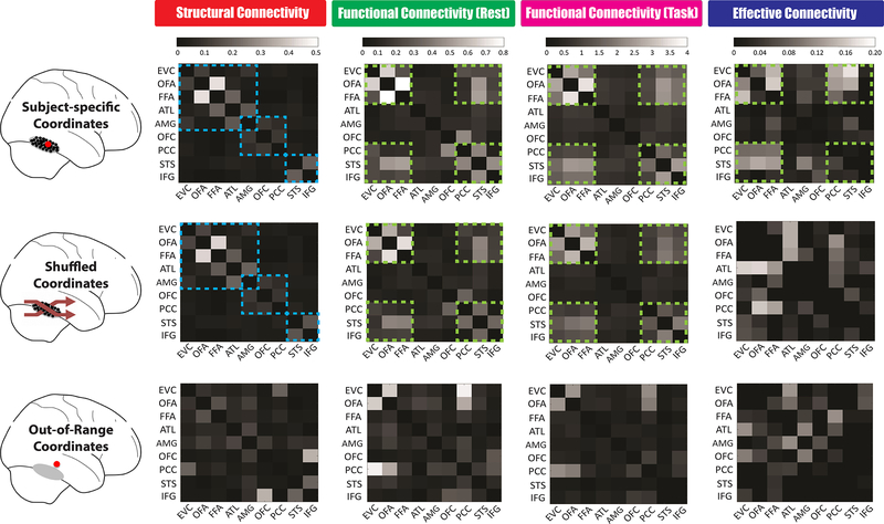
Figure 5. The Spatial Specificity of Face Connectome Organizations in the Right Hemisphere.
Brain connectivity patterns were measured and compared across three different ways of ROI definition. We first (Top Row) defined each ROI by using subject-specific coordinates from the functional face localizer. The major findings in this article came from this method, such as three distinct pathways founded in the structural connectivity (blue dash line) and six synchronized areas exhibited in the functional and effective connectivity (Green dash line). We then (Middle Row) permuted subject-specific coordinates across subjects (e.g. subject A’s FFA coordinates now became subject B’s; subject C’s ATL coordinates now became subject D’s). Since the individual variations of each ROI’s coordinates can be typically circumscribed by a 10–15mm radius sphere (see group coordinate ranges listed in Table S1), these new shuffled coordinates were in relatively close proximity to the original subject-specific ones. We found the patterns of structural connectivity and functional connectivity during rest and task remained the same after this permutation manipulation but the effectivity connectivity was changed significantly. Lastly (Bottom Row), we chose each ROI even farther away in space—with new coordinates just outside the putative range (i.e. 20 mm apart from its original coordinates, so that subject A’s FFA coordinates could now fall into inferior temporal gyrus). After this 20mm shifting manipulation, these out-of-range coordinates drastically altered all four brain connectivity patterns, thus showing spatial specificity of our findings. Similar results of spatial specificity were found in the left hemisphere.

