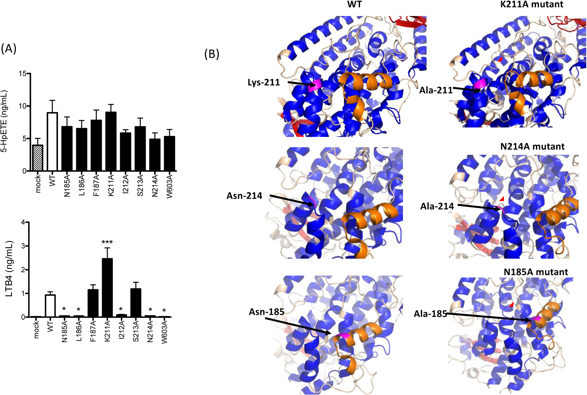
Figure 3. 5-lipoxygenase activity of mutants at pocket 1.

(A) Alanine mutants were transiently transfected into HEK cells as described in the Method. 5-lipoxygenase activity of alanine mutants was measured. Data were shown as mean +/− S.D. of 4 replicates. Two independent experiments were performed. Statistical analysis was performed using one-way ANOVA with Bonferroni post hoc analysis. * and *** denote p< 0.05 and p< 0.001, respectively.
(B) Predicted structures of alanine mutants were shown. Magenta showed residues to be mutated or mutated. Orange showed the α2 helix. Red circle highlighted the structural change of the α2 helix.
