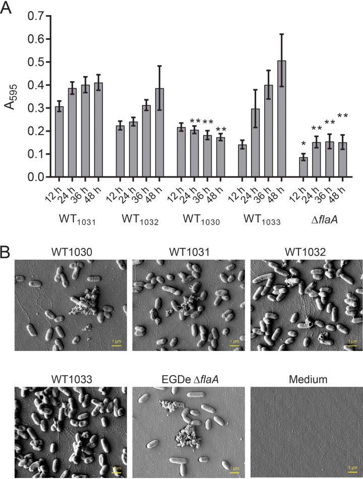
FIG 3.
Biofilm formation of the four L. monocytogenes EGDe isolates. (A) The biomasses of the four EGDe isolates adherent to the substratum were quantified over time when incubated at 30°C. The EGDe ΔflaA strain was used as a negative control. The values presented are the means from 29 independent experiments for the EGDe isolates and 4 experiments for the ΔflaA strain. The error bars are the standard errors of the means. The data were analyzed by one-way ANOVA comparing with WT1031. *, P ≤ 0.05; **, P ≤ 0.01. (B) The biomass adherent to the substratum was imaged using scanning electron microscopy after 48 h of incubation. The representative images shown were taken at the midpoint of the peg.

