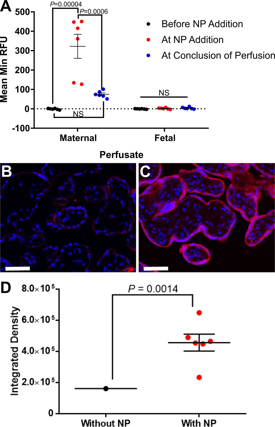
Figure 1. Analysis of Texas-red fluorescence in maternal and fetal perfusate samples, and placental tissue from the dual placental perfusion experiments.

Prior to addition of Texas-red conjugated nanoparticle (NP) to maternal perfusate, no Texas-red fluorescence was recorded in maternal perfusate (A). Upon addition of nanoparticle, Texas-red fluorescence increased in maternal perfusate and declined after approximately 1 hour of perfusion. Texas-red fluorescence was not determined in fetal perfusate at any of the time points (A). Histological analysis of placental tissue collected at the conclusion of the experiment showed nanoparticle localization in the syncytiotrophoblast layer of the fetal villi (B negative control tissue: perfused tissue with no Texas-red conjugated nanoparticle added, C positive tissue: tissue perfused with Texas-red conjugated nanoparticle for approximately 1 hour). Fluorescence analysis of the tissue sections confirmed a significant increase in Texas-red in the tissue sections exposed to nanoparticle (D). Data are mean ± SEM, n=7 term placentas perfused; n=6 term placentas perfused with NP. Statistical significance determined with an ANOVA and Tukey’s post hoc analysis (A) and Mann-Whitney test (D). Scale bar = 50 μm.
