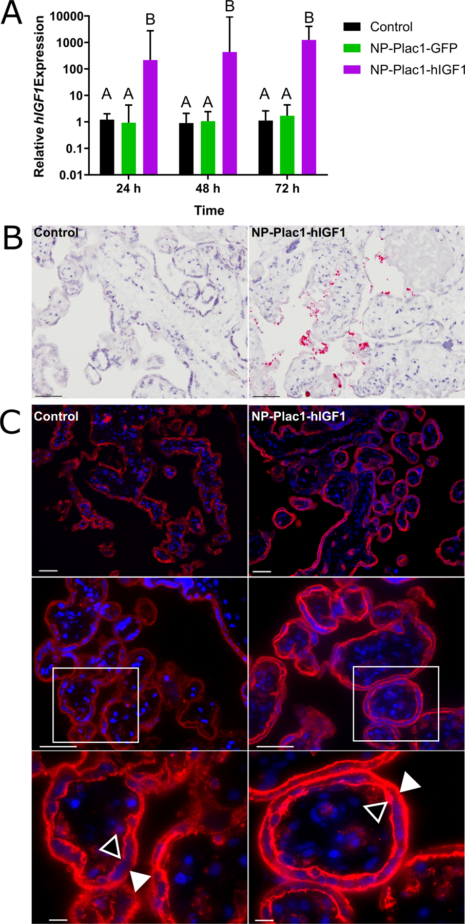
Figure 3. qPCR analysis of human insulin-like 1 (hIGF1) mRNA expression and immunohistochemical (IHC) localization of glucose transporter 1 (SLC2A1) in placenta fragments treated with nanoparticle.

qPCR analysis of hIGF1 mRNA showed a significant increase in placenta fragments treated with nanoparticle (NP-Plac1-hIGF1) at 24 hours which was sustained at 72 hours (A). There was no increase in hIGF1 mRNA expression in fragments treated with nanoparticle containing a plasmid encoding the green fluorescent protein gene (NP-Plac1-GFP)(A). In situ hybridization confirmed plasmid specific mRNA expression of IGF1 in syncytiotrophoblast cells of fragments 72 h after treatment with NP-Plac1-hIGF1 and not in untreated fragments (B). Representative images of IHC staining of SLC2A1 in fragments at 48 hours showed trans-localization of the transporter to the apical (closed triangle) and basal (open triangle) membranes of the syncytiotrophoblast and cytotrophoblasts cells in the fragments treated with nanoparticle (NP-Plac1-hIGF1) compared to untreated (control) (C). Data are median ± interquartile range, n=6 term placentas. Scale Bar: 200 μm (C top row), 50 μm (B and C middle row), 10 μm (C bottom row). Statistical significance was determined with a 2-way ANOVA.
