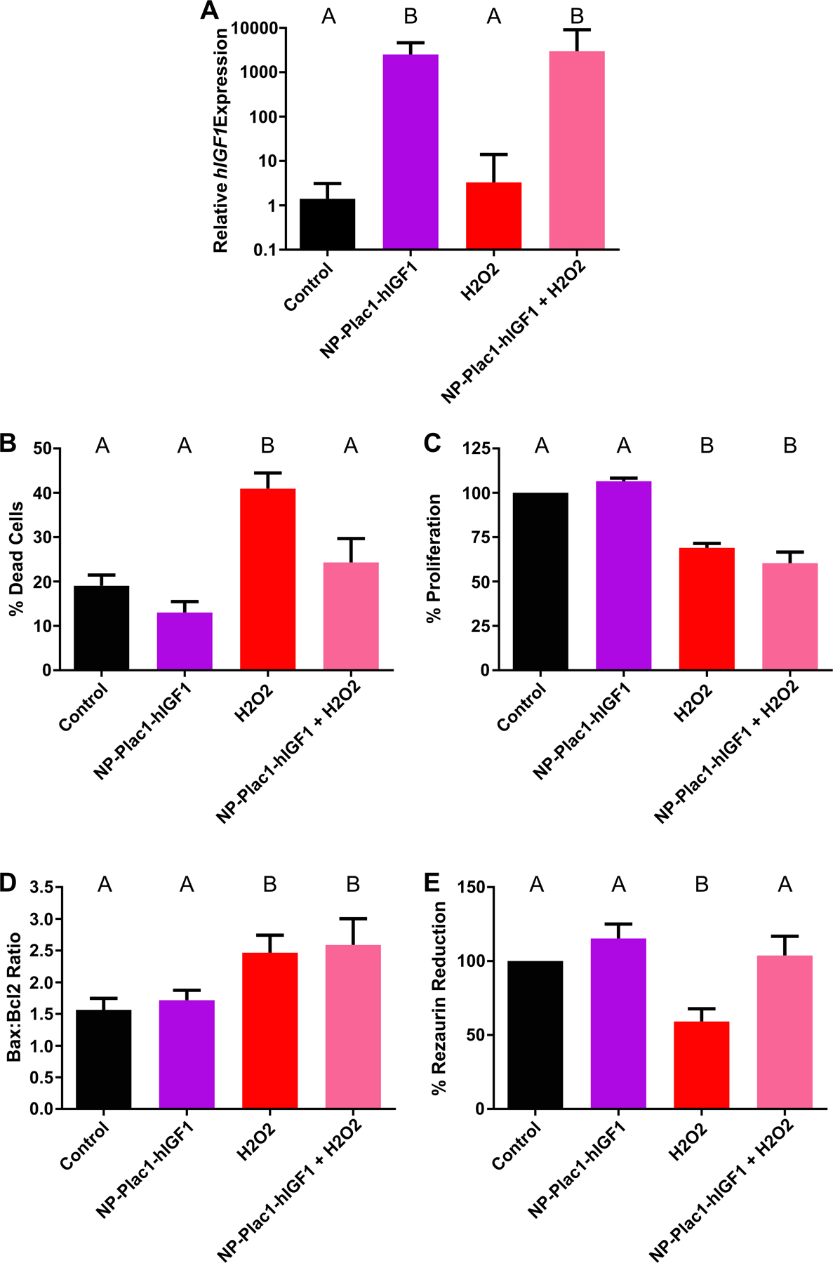
Figure 4. Functional effect of nanoparticle treatment in protecting against oxidative stress in BeWo cells.

qPCR analysis of human insulin-like growth factor 1 (hIGF1) was increased in cells treated with nanoparticle (NP-Plac1-hIGF1 and H2O2+NP-Plac1-hIGF1) compared to untreated (control) and H2O2 alone (A). Compared to untreated, treatment with H2O2 significantly increased the percentage of dead cells but not when cells were pretreated with nanoparticle (B). Cell number was lower in cells treated with H2O2 alone and nanoparticle with H2O2 compared to untreated and nanoparticle alone treated (C). The BAX:BCL2 ratio was higher in cells treated with H2O2 as well as in those treated with nanoparticle and H2O2 when compared to untreated and nanoparticle alone treated (D). Mitochondrial activity, as measured by the percentage of rezaurin reduction, was lower in cells treated with H2O2 alone compared to untreated but not in cells treated with nanoparticle prior to H2O2 treatment (E). Data are median ± interquartile range (A) or mean ± SEM (B-E), n=6 passages. Statistical significance was determined with a repeated measures ANOVA and Sidak’s multiple comparison test.
