
Figure 4.
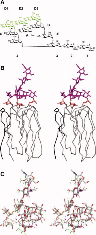
Modeling of the interactions of Man9GlcNAc2 with griffithsin. (A) Chemical structure of Man9GlcNAc2, with the three terminal mannose residues that make direct contact with griffithsin colored green. In the resulting model described here, mannose D1 is equivalent to Man11, D2 to Man8, and D3 to Man6. (B) Chain tracing of a single domain of griffithsin, consisting of residues A1–A18 (black) and B19–B121 (gray), together with the model of bound Man9GlcNAc2. The latter is colored in magenta, except of the three terminal residues that are colored in gold. (C) Superposition of the coordinates of the model of Man9GlcNAc2 complexed with griffithsin (colored sticks) with the experimentally determined structures of Man9GlcNAc2 in complex with the specific antibody 2G12 25 (thin green and magenta lines).