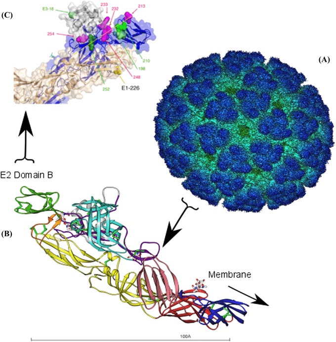
Figure 1.

Alphavirus infection of host cells is mediated by two viral glycoproteins, E1 and E2. In this figure, the chikungunya virus cryoelectron structure is used to illustrate the location of E2 domain B, an immunodominant region of E2 where host range–expanding mutations are commonly found. (A) The cryoelectron structure of the entire icosahedral chikungunya virus particle is shown. The E1 and E2 glycoproteins dimerize and form 80 membrane‐embedded trimeric spikes across the virus capsid surface, shown in dark blue. E2 binds the host cell laminin receptor, and E1 induces the fusion of the viral and host cell membranes, allowing the nucleocapsid to enter the cell. Figure downloaded from Protein Data Bank Japan (PDBj). (B) A ribbon diagram shows the E1–E2 heterodimer. Each E1–E2 heterodimer is aligned so that E2 domain B (shown in green) is at the membrane distal end, and the opposite end (E1 domains I and III in red and blue, respectively) is closest to the virus lipid bilayer. Figure courtesy of Marie‐Christine Vaney and Félix Rey, Institute Pasteur; reprinted from Ref. 24. (C) A close‐up of E2 domain B (blue) shows the locations of substitutions (pink and green spheres) that increase chikungunya virus fitness in A. albopictus (except for a T213Q substitution, see Ref. 38). Figure modified from Ref. 38.
