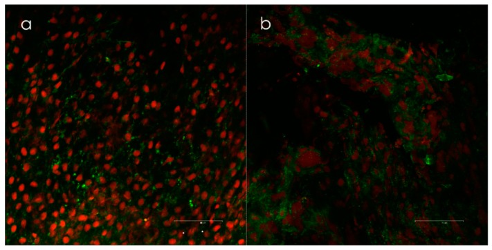
Figure 3.
(a) Biofilm positive samples taken from the lateral side of concha media visualized by confocal scanning laser microscopy (Leica TCS SP2 AOBS) and (b) live/dead-staining (Invitrogen’s LIVE/DEAD BacLight™, Invitrogen, Burlington, Canada). Epithelial cells are red, and the bacteria are green. Biofilms were scored when clusters of bacteria with intact membranes were present in both the x-y and x-z axes. Courtesy of Dr. Kjell Arild Danielsen.

