
Figure 1.
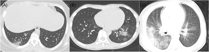
A, Female, 14 years old. Chest CT showed scattered ground‐glass opacities in the inferior lobe of the right lung, located subpleural or extended from subpleural lesions. B, Male, 10 years old. Chest CT showed consolidation with halo sign in the inferior lobe of the left lung surrounded by ground‐glass opacities. C, Male, 1 year old. Chest CT showed diffused consolidations and ground‐glass opacities in both lungs, with a "white lung" appearance of the right lung. CT, computed tomography