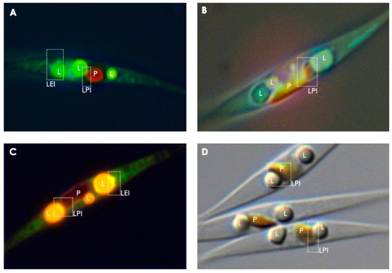
Figure 2.
Micrographs of nitrogen starved P. tricornutum cells, illustrating potential interconnectivity between LDs and various cellular compartments. Plastidial autofluorescence appears red, the LD stain Nile Red fluoresces yellow and the ER/mitochondrial/endomembrane stain DiOC6 fluoresces green/greenish blue. (A) Epifluorescent image of cells stained with Nile Red and DiOC6, (B) Epifluorescent image of cells stained with only DiOC6, (C) Epifluorescent image of cells stained with Nile Red and DiOC6, (D) Differential interference contrast image with no epifluorescent staining. P = plastid, L = lipid droplet, LPI = lipid droplet-plastid interface, LEI = lipid droplet-endomembrane interface. The interfacial regions, emphasized in boxes with dotted lines, are speculated to be potential regions of interaction between LDs and other organelles.

