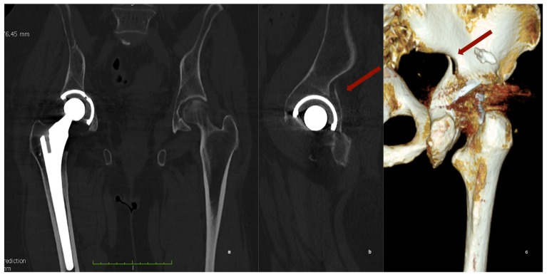
Figure 2.
CT scan of the pelvis. (a) Coronal view shows a slightly medially protruded acetabular cup; (b) the sagittal view of the hip, revealed the posterior wall fracture of the acetabulum and (c) in the tridimensional reconstruction the fracture is clearly visible, but its extension is hidden by image artifacts.

