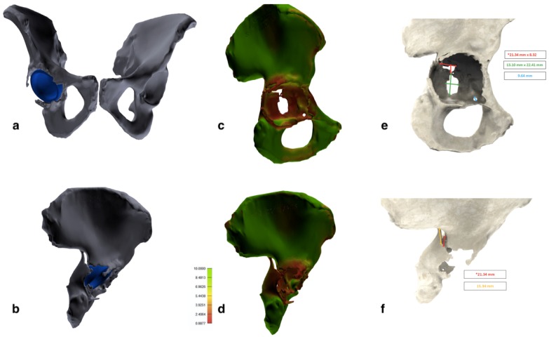
Figure 3.
Tridimensional images elaborated with the 3D modeling software. (a,b) Entire pelvis with the acetabular cup retained. Femurs and femoral stem were removed during segmentation. (c,d) Bone quality map shows regions with normal bone quality (green) and regions with low bone quality and thickness (red). (e,f) Measurements of the bone defect area and fracture extension.

