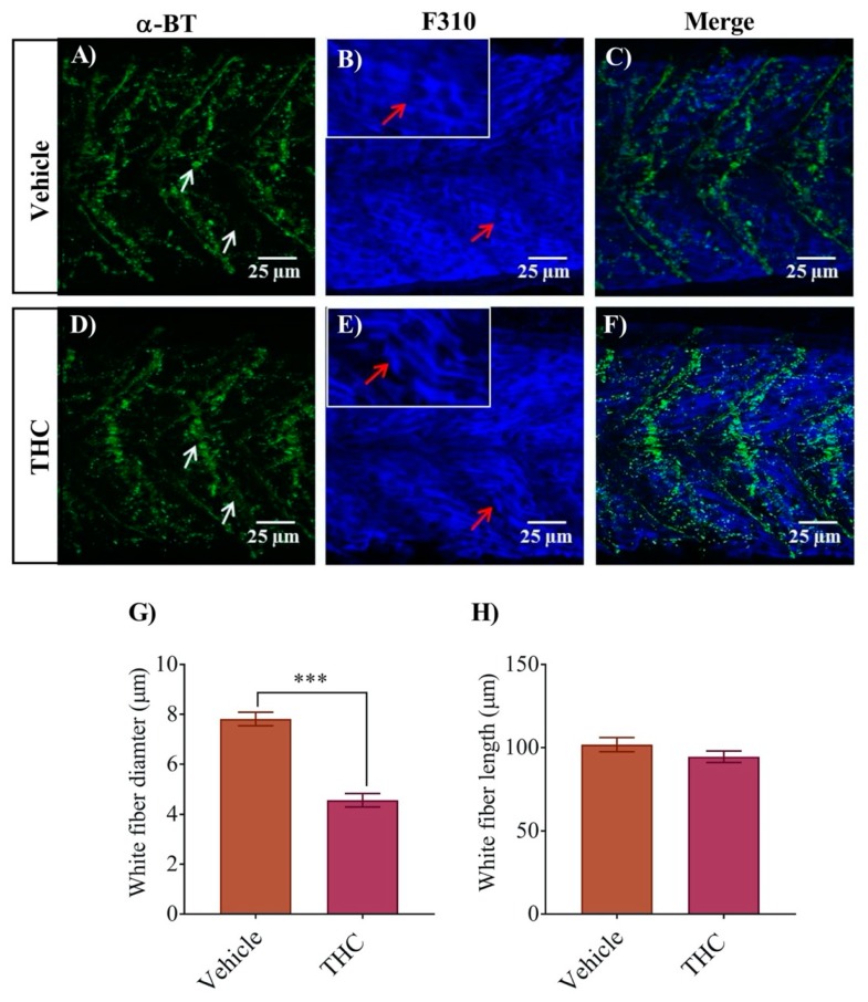
Figure 4.
Co-labeling of white muscle fibers and nAChRs using anti-F310 and Alexa 488 conjugated α-bungarotoxin respectively. (A) α-Bungarotoxin labelled nAChRs associated with white muscle fibers in vehicle-treated embryos. White arrow shows clusters of nAChRs. (B) Anti-F-59 labelled muscle fibers. Red arrows point to the edge of a muscle fiber. Inset shows muscle fibers at higher magnification to better determine the size of the fiber. (C) Merged image showing the co-labeled white muscle fiber and nAChR in vehicle-treated animals. (D) α-bungarotoxin labelled nAChRs associated with white muscle fibers in THC-treated (6 mg/L) embryos. White arrow shows clusters of nAChRs. (E) Anti-F310 labelled muscle fibers. Red arrows point to the edge of a muscle fiber. Inset shows muscle fibers at higher magnification to better determine the size of the fiber. (F) Merged image showing the co-labeled white muscle fiber and nAChR in THC-treated animals. (G) Bar graph showing the diameter of white fibers for vehicle and THC treated embryos and (H) Measurement of white fiber length. *** significantly different from vehicle control, p < 0.001.

