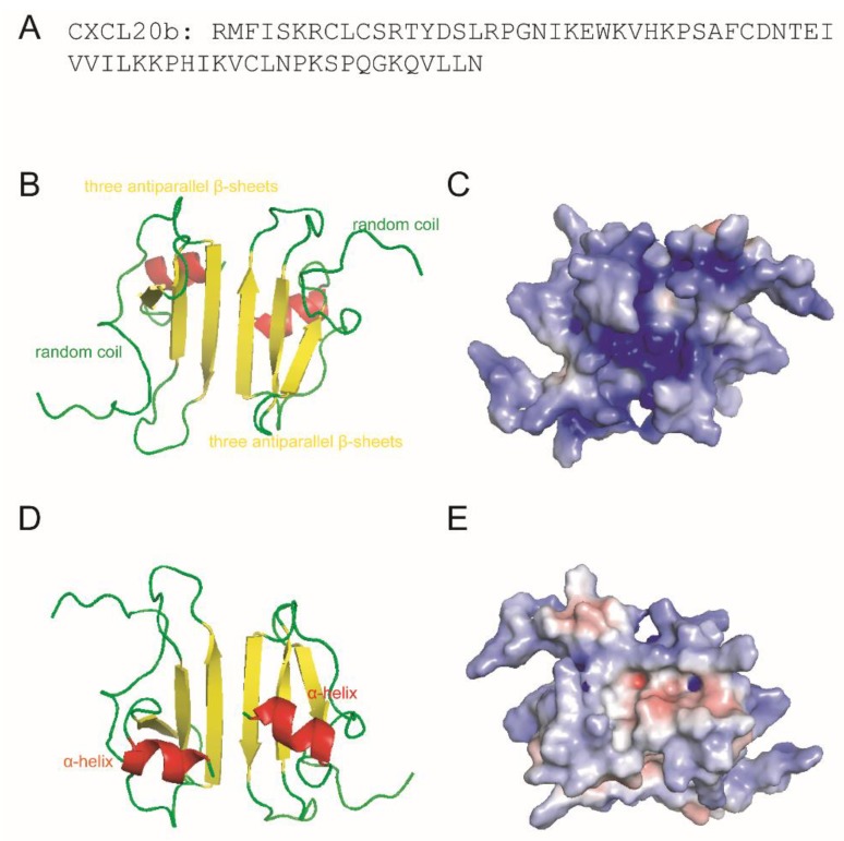
Figure 2.
3D modeling and surface charge distribution analysis of grass carp CXCL20b. (A) The amino acids sequence of CXCL20b. (B,D) On the basis of the prediction of grass carp CXCL20b, the images show a 3D model of CXCL20b dimers. Grass carp CXCL20b features a typical mammalian chemokine structure, including N-terminal random coil (green), core structure containing three antiparallel β-strands (yellow), and C-terminal α-helix (red). (C,E) Electrostatic potentials mapped onto the surfaces of grass carp CXCL20b. Blue represents the cationic regions, red represents the negative regions, and white represents the hydrophobic residues.

