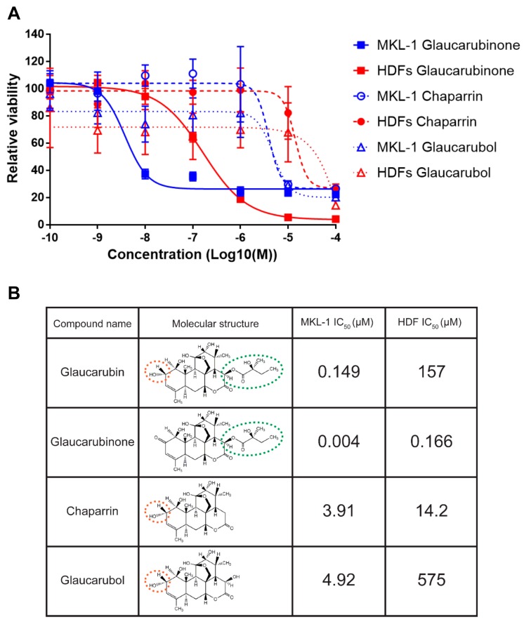
Figure 2.
Glaucarubin derivatives inhibit the growth of MCC and HDF cells. (A) Cell viability assay of MCPyV-positive MCC cell line MKL-1 (blue) and HDFs (red) treated with the indicated concentrations of glaucarubin analogs 72 h post-treatment. (B) Table highlighting the IC50s of each compound from (A) in MKL-1 and HDF cells. Molecular structures of each compound are included for the purpose of relating features of the compounds to phenotypes in cells. For example, the ester-linked moiety featured on the lactone of glaucarubin (circled with a green dotted line) appears necessary for the cytotoxicity in MKL-1 cells, while the hydroxyl group (circled with an orange dotted line) results in less cytotoxicity in HDFs than a carbonyl group at the same position.

