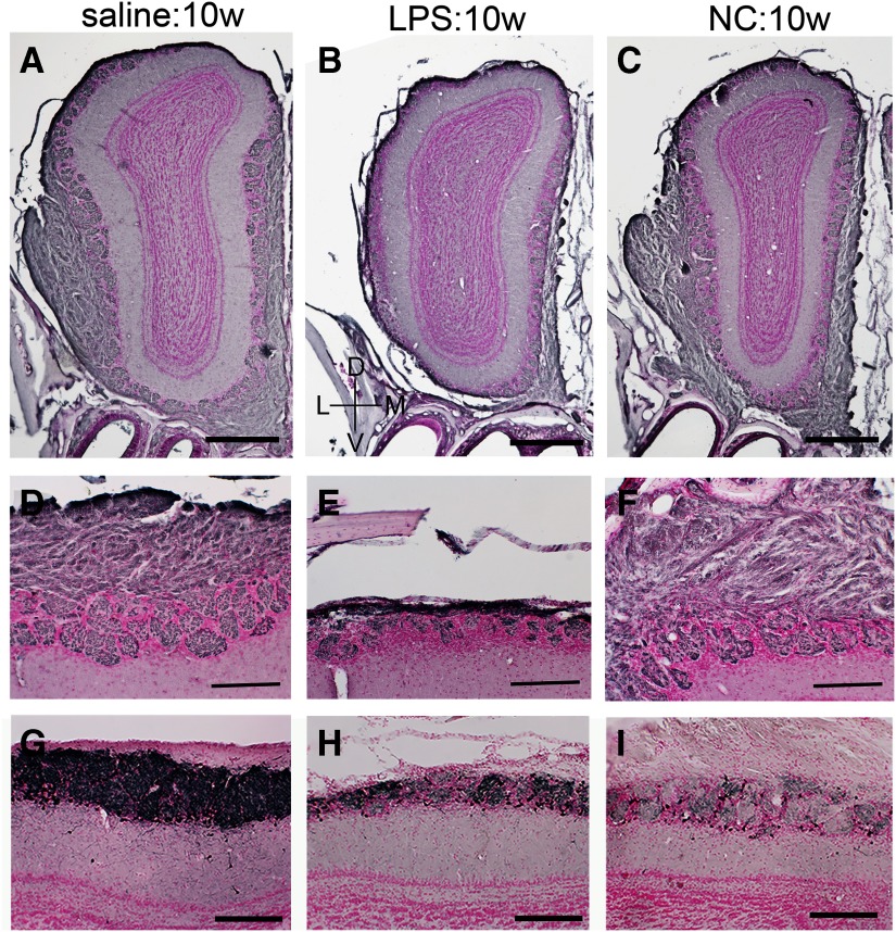
Figure 4.
OMP and TH expression in the OB. A–F, Immunohistochemistry for OMP, representing ONL and GL, in ipsilateral OBs from saline:10w (A, D), LPS:10w (B, E), and NC:10w (C, F). OMP-immunopositive axon terminals of OSNs were lost particularly in the lateral side of the ipsilateral OB in LPS:10w (B, E) but not in saline:10w (A, D) or NC:10w (C, F). D–F, Magnified views of lateral side of the OBs in A–C. G–I, Immunohistochemistry for TH expressed by some juxtaglomerular cells. TH expression decreased in LPS:10w (H) and in NC:10w (I) compared with that in the saline:10w (G). Nuclei were stained with nuclear fast red. Scale bars: 500 μm (A–C) and 200 μm (D–I).

