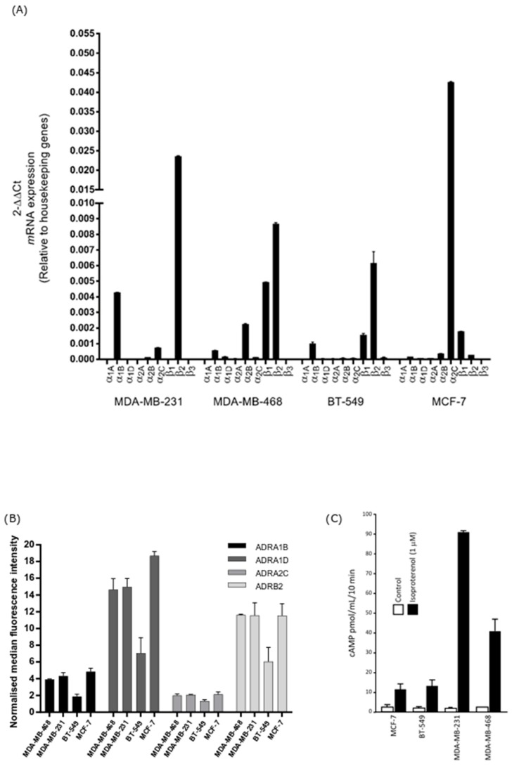
Figure 1.
Adrenoceptor (ADR) expression on breast cancer cells and measurement of cAMP (cyclic adenosine monophosphate) levels following adrenergic stimulation. (A) Relative expression of ADR mRNA in breast cancer cell lines was quantified by qRT-PCR (quantitative reverse transcription polymerase chain reaction) and relative expression (2−ΔΔCT) was determined by normalisation to housekeeping genes. β2-adrenoceptor gene expression was strongest in the basal-type cell lines MDA-MB-231 and MDA-MB-468. (B) Expression of the adrenoceptors on unpermeabilised breast cancer cells was assessed by measuring median fluorescence intensity (MFI) using flow cytometry. Membranous β2-adrenoceptor protein expression was highest in the basal-type cell line MDA-MB-468, followed by MDA-MB-231 > MCF-7 > BT-549. (C) Isoproterenol (β-agonist) stimulated cAMP accumulation in breast cancer cell lines. Cells were treated in the presence of IBMX to prevent cAMP degradation. cAMP production was highest in the order MDA-MB-231 > MDA-MB-468 > BT-549 > MCF-7. All assays were performed in triplicate (n = 3). Results shown are the mean ± standard deviation.

