
Figure 4.
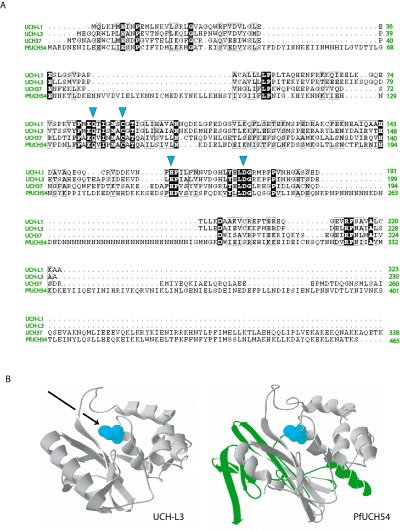
Alignment of PfUCH54 with human DUB homologues and predicted structure. A. An alignment of three human UCH‐type DUBs, UCH‐L1, UCH‐L3 and UCH37, with PfUCH54 is shown with active residues indicated by the blue arrows. B. The crystal structure of UCH‐L3 was used to model PfUCH54. Parasite‐specific insertions are shown in green. Active site cysteine residues for both enzymes are shown in blue. The arrow indicates the trajectory of Ub that is presumed for binding to UCH‐L3.