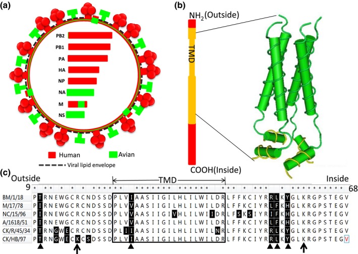
Figure 4.

Schematic diagram of the Spanish flu virus with the recombinant M segment. (a) The schematic diagram of the Spanish flu virus structure. Segments of different origins are indicated with different colours. The encoded proteins are described using the identical colour respectively. The recombinant M segment is shown with two different colours, red and green. (b) The schematic diagram of the mosaic M2 protein of the Spanish flu virus and the 3D structure diagram of its pH‐gated proton channel encoded by genetic materials originating from avian influenza virus. TMD indicates the region of transmembrane domain, and is framed in the box. The amino acids (AAs) in the inside helix are shown in yellow. The 3D structure of the proton channel is deviated from the previous report (PDB ID: 2RLF). (c) The AA comparison of the Spanish flu virus and its parent lineage representatives in the recombination region of M2. The asterisks indicate the identical AAs between the Spanish flu virus and its parents. The triangles show the unique AAs of bird lineage and the Spanish flu virus. The two black arrows indicate the start and end points of the pH‐gated proton channel. The AAs masked with light yellow constitute the inside helix of the channel. BM/1/18, A/Brevig_Mission/1/1918(H1N1); NC/15/97, A/Nanchang/15/1996(H1N1); M/17/78, A/Memphis/17/1978(H1N1), A/1618/51, A/Albany/1618/1951(H1N1) CK/R/45/34, A/chicken/Rostock/45/1934(H7N1); CK/HB/97, A/chicken/Hubei/wi/1997(H5N1) [Colour figure can be viewed at http://wileyonlinelibrary.com]
