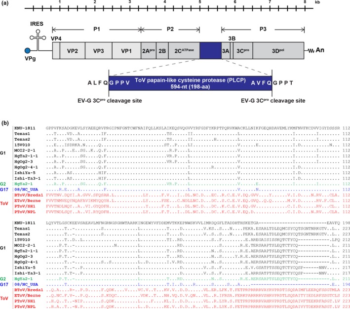
Figure 1.

Schematic diagram of the genome organization of KOR/KNU‐1811/2018/G1‐PLCP. (a) The Korean KNU‐1811 genome contains a single open reading frame (ORF) flanked by a long 5′ untranslated region (UTR) (813‐nt) and a short 3′ UTR (71‐nt), followed by a poly (A) tail (An). The stem‐loop secondary structure on the 5′ UTR represents an internal ribosome entry site (IRES). The torovirus (ToV) papain‐like cysteine protease (PLCP) gene is presented as a blue box that is inserted at the 2C/3A cleavage junction. The 5′‐ and 3′‐boundary sequences of enterovirus (EV‐G) 3C protease (3Cpro) cleavage sites are shown in an enlarged blue box. Vertical lines indicate the polyprotein processing sites creating precursor polyproteins P1, P2, and P3 by 3Cpro. (b) Multiple alignment of the amino acid sequences of the PLCP regions of the recombinant EV‐G and ToV strains [Colour figure can be viewed at http://wileyonlinelibrary.com]
