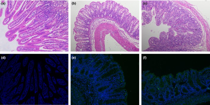
Figure 9.

Microscopic small intestine lesions in piglets inoculated with PEDV HLJBY strain. (a) Small intestines from a mock piglet. The normal villous was observed in the mock‐inoculated piglet. (b) Small intestines from a piglet inoculated with PEDV CV777 strain, showed villous atrophy. (c) Small intestines from a piglet inoculated with PEDV HLJBY strain showed diffuse atrophic enteritis. (d–f) Detection of PEDV N antigen in the small intestines was performed using immunofluorescence (100× magnification). PEDV N was green (FITC) and DAPI was blue. (d) No PEDV N antigen was detected in the small intestines of the mock‐inoculated piglet. (e–f) Immunostaining of PEDV N antigen was detected in the epithelial cells of the small intestines in PEDV CV777 (e) and HLJBY (f) inoculated piglets [Colour figure can be viewed at http://www.wileyonlinelibrary.com/]
