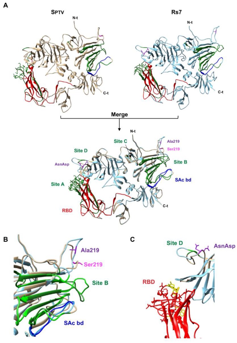
Figure 5.
Comparison of SPTV and Rs7 structure models. (A) The S1 domains (aa 1 to 818) from SPTV (left panel, light brown) and Rs7 (right panel, light blue) were modelled and compared (merged structures in the bottom panel). Relevant domains are indicated, such as pAPN (receptor) binding domain (RBD, red), sialic acid binding domain (SAc bd, blue), and the antigenic sites A, B, C, and D (green). Site A partially overlaps with RBD. Ser219, present only in SPTV sequence, is indicated in magenta. Ala219 and the Y374_T375insND (AsnAsp), present only in the engineered Rs7 sequence, are indicated in purple. (B) Detail of the region around Ser219. Labels and colors as in panel A. (C) Detail of the region around the ND insertion in Rs7. The RBD residues involved in S-pAPN contacts are indicated, including those engaged in hydrogen bonding (in yellow) [10]. Labels and colors as in panel A.

