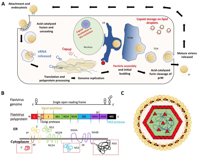
Figure 1.
(A) Diagram of flavivirus life cycle with emphasis on distribution of viral capsid (red). (B) Schematic of the flaviviral genome, polyprotein, and transmembrane viral proteins. Adapted from Ming et al. [3]. Red boxes indicate soluble viral proteins in the cytoplasm. The primary focus of this review will be the viral capsid [C] protein. (C) Diagram of flavivirus particle with E (yellow), M (orange), and C (red) proteins. E and M proteins span the membrane derived from host endoplasmic reticulum and capsid interacts with these proteins as well as coats the viral genome (blue line).

