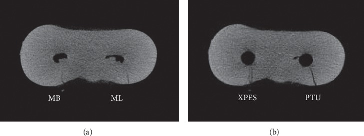
Figure 3.

Micro-CT cross-sectional image of the cervical third of mandibular molar mesial root: (a) Preinstrumentation; (b) postinstrumentation. The MB canal (left) instrumented with XPES and the ML canal (right) instrumented with PTU. The microcracks that appeared in the postinstrumentation image for both canals were the propagation of previous microcracks observed in the preinstrumentation image.
